


Servicios Personalizados
Revista

Articulo
 Inglés (pdf)
 Articulo en XML
 Referencias del artículo
 Enviar articulo por email
Indicadores
-
 Citado por SciELO
Links relacionados
-
 Similares en
SciELO
Similares en
SciELO
Compartir

 Permalink
Acta Odontológica Latinoamericana
versión On-line ISSN 1852-4834
Acta odontol. latinoam. vol.23 no.3 Buenos Aires dic. 2010
ARTÍCULOS ORIGINALES
Effect of bleaching agent on dental ceramics roughness
Aleska D Vanderlei, Sheila P Passos, Susana M Salazar-Marocho, Sarina Mb Pereira, Vanessa Zc Vásquez, Marco A Bottino
Department of Dental Materials and Prosthodontics, São Paulo State University at São José dos Campos, Brazil.
CORRESPONDENCE Aleska Dias Vanderlei Rua Ibituruna, 104, ap 131, saude, Sao Paulo SP CEP 04302050, Brasil E-mail: aleskavanderlei@hotmail.com
ABSTRACT
The aim of this study was to assess the effect of bleaching agents (10% and 16% carbamide peroxide) on the roughness of two dental ceramics in vitro, and to analyze the surface by scanning electronic microscopy (SEM). Two bleaching agents (10% and 16% / Whiteness, FGM Gel) and two microparticle feldspathic ceramics (Vita VM7 and Vita VM13) were used. Forty disks of Vita VM7 and Vita VM13 ceramic were manufactured, measuring 4mm in diameter and 4mm high, in accordance with the manufacturers’ recommendations, and were divided into 4 groups (n=10): (1) VM7 + Whiteness 10%; (2) VM7 + Whiteness 16%; (3) VM13 + Whiteness 10%; (4) VM13 + Whiteness 16%. The bleaching agent was applied for 8 hours a day for 15 days and during the intervals the test specimens were stored in distilled water at 37°C. The roughness (Ra) of the test specimens was evaluated before and after exposure to the bleaching agents using a laser roughness meter and the topographic description was analyzed by SEM. The statistical analysis of roughness data showed significant differences in the VM7 groups, using paired t-test, p=0.05 (VM7 + Whiteness 10%: p=0.002; VM7 + Whiteness 16%: p=0.001) and two-sample t-test (VM7 p=0,047), and no significant difference was found among VM13 groups. The qualitative SEM analysis showed different degrees of surface changes. The results suggest that the roughness of the tested ceramic surfaces increased after exposure to the bleaching agents.
Key words: Ceramic; Roughness; Bleaching agents.
RESUMO
Efeito dos agentes clareadores na rugosidade da cerâmica dental
O objetivo do estudo foi avaliar o efeito de agente clareador (10% e 16% de peroxido de carbamida) na rugosidade de duas ceramicas dentais in vitro e analisar a superficie atraves da microscopia eletronica de varredura (SEM). Foram utilizados dois agentes clareadores (10% e 16% / Whiteness, FGM Gel) e duas ceramicas feldspaticas microparticuladas (Vita VM7 e Vita VM13). Foram confeccionados quarenta discos de ceramica Vita VM7 e Vita VM13, com 4mm de diametro e 4mm de altura, de acordo com as recomendacoes do fabricante, sendo divididos em 4 grupos (n=10): G1- VM7 + Whiteness 10%, G2- VM7 + Whiteness 16%, G3- VM13 + Whiteness 10% e G4- VM13 + Whiteness 16%. O agente clareador foi aplicado por 8 horas durante 15 dias e durante os intervalos os corpos de prova (cp) ficavam armazenados em agua destilada a 37°C. A rugosidade (Ra) dos cp foi avaliada antes e apos a exposicao aos agentes clareadores utilizando o rugosimetro a laser Perthomether S8P, e a descricao topografica foi analisada no MEV. A analise estatistica na rugosidade mostrou diferenca significante entre os grupos da VM7, usando o teste pareado, p=0.05 (VM7 + Whiteness 10%: p=0.002; VM7 + Whiteness 16%: p=0.001) e nao foi encontrado diferenca estatistica para os grupos da VM13. A descricao qualitativa no SEM evidenciou alteracao nas superficies em diferentes graus. Os resultados sugeriram que as superficies das ceramicas testadas aumentaram a rugosidade apos a exposicao ao agente clareador.
Palavras chave: Ceramica; Rugosidade; Agente clareador.
INTRODUCTION
Dental bleaching is a procedure that has become increasingly popular in dentistry because it appears to be efficient and non-invasive. The treatment can be performed in the dental office by the dentist, or at home by the patient himself 1,2. Among the agents available, there are carbamide peroxide (CP) gels at concentrations between 10-40%. Bleaching times vary according to the gel concentration and technique used3. Carbamide peroxide is an oxidizing agent; consisting of hydrogen peroxide compounded with urea, that when it comes into contact with the oral tissues and saliva, its constituent parts separate3. The 10% to 16% CP solution dissociates into 3% to 5% hydrogen peroxide and 7% to 10% urea. The hydrogen peroxide further degrades into oxygen and water, whereas the urea degrades into ammonia and carbon dioxide3,4. It is believed that dental bleaching occurs due to modifications in the chemical structure of organic substances, by free radicals that are generated by the bleaching agents 5, by oxidation reaction2,6. However, due to the close contact between the bleaching agent and restorative materials, it is important to reflect on the negative alterations caused on them7-9.
Although dental bleaching does not cause macroscopically visible alterations, microscopic alterations may cause undesirable effects. Many studies have assessed the influence of bleaching agents on oral tissues, enamel and dentin10-20. Some studies have assessed the effects of bleaching agents on composites8,21- 27. However, there are hardly any reports on the effects of dental bleaching on dental ceramics, which have been an excellent alternative to substitute metal restorations, due to their characteristics of safety and efficiency when adequate indications and techniques are used. Several authors25,28,31 evaluated the effect of bleaching agents on the surface texture of porcelains. In the study by Butler et al.31, porcelains showed a significant increase in roughness data when exposed to 10% CP and the roughness size obtained was approximately 0.24 μm, which, according to the authors, might increase plaque accumulation or affect the esthetics by changing the texture of the ceramic restoration. Studies on the effect of CP gels on the surface roughness of ceramic dental materials after different exposure times have reported some conflicting results. Moraes et al.28 reported that when feldspathic porcelain is exposed to both home and in-office bleaching agents, the porcelain surface roughness increased after 21 days of exposure. Ceramics occupy an outstanding position because of their optic properties, which are similar to those of natural teeth, physical-mechanical durability, chemical stability and optimum biocompatibility with the adjacent tissues and low biofilm adherence index29,30 as a result of the highly smooth surface obtained with the new ceramics. Nevertheless, there are factors that may influence the quality of a restoration, such as the oral environment, bacteria, saliva, stress, habits, cleaning products, mouthwashes and others that undoubtedly contribute to deterioration of the restoration and may necessitate restoration replacement29. Clinically, surface integrity and smoothness are important factors when the restoration is evaluated, as they are related to bacterial plaque34, abrasion of antagonistic teeth, injury to adjacent soft tissues, color changes and also to the esthetic quality of the restoration6,9,14,23. The clinical implications also include the decrease in the flexural strength of the porcelain materials35.
In some studies the quantitative results are obtained by means of the roughness25,28,31; however, the statistical results should be verified with scanning electron microscopy (SEM) images to evaluate the surface topography, allowing a definition of form and contour, which are not always detected by the filters of the surface roughness tester. SEM is used for characterizing surface topography. SEM images have great depth of field, yielding a characteristic three-dimensional appearance useful for understanding the surface structure of a sample. This great depth of field and the wide range of magnifications are the most familiar imaging mode for specimens in the SEM. Thus, all the studies about roughness used SEM to determine changes in surface morphology8,25,36-38. Considering the negative effects of the bleaching agents on the restorative material, these effects may result in changes in physical properties, surface morphology and color of different restorative materials21. The aim of this study was to assess the surface alteration of ceramic materials on exposure to 10% and 16% CP for 15 days in vitro, by means of surface roughness and topographical analyses by SEM. The null hypotheses to be tested are that: (1) 10% CP bleaching agent does not influence the roughness of VM7 and VM13 ceramic surface; and (2) 16% CP bleaching agent does not influence the roughness of VM7 and VM13 ceramic surface.
MATERIALS AND METHODS
Forty disks were prepared with the ceramic materials listed in Table 1. All samples were handled in accordance with the manufacturer’s directions. A hollow, cylindrical 4x4 mm (diameter and height) metal mould was used as standard.
Table 1: Information on materials used in the study, according to manufacturers.

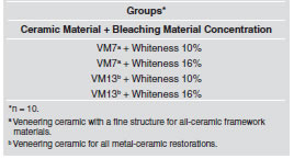
The Vita VM7 and Vita VM13 ceramics were prepared by adding the modeling liquid to the powder until a creamy consistency was obtained. The pastes were put into the mould with a TeflonR spatula to model them, and excess water was eliminated with absorbent paper. After modeling each set of 5 ceramic paste disks, the disks were sintered in a Vacumat 40 vacuum (Vita, Zahnfabrik-Germany), according to manufacturer’s recommendations, from 500oC to 910oC in 6 minutes in vacuum with a 1-minute hold at peak temperature. After firing, the specimens were included in acrylic resin and finished with a polishing machine (Labpol 8-12, Extec, USA), using sandpaper No 600, 800 and 1200 (3M, St. Paul, USA) to remove any irregularities and to create a flat surface. All the ceramic disks were cleaned in an ultrasonic bath for 5 min with distilled water. Forty disks were randomly divided into 4 experimental groups, according to the ceramic (VM7 and VM13) and the bleaching agent concentration (10% and 16% / Whiteness, FGM Gel) (Table 2).
Table 2: Experimental groups.
The surface roughness (Ra) was determined using a laser roughness meter Perthomether S8P with a T9 Fododyn tip, optical micro palpation instrument from IEAv/CTA. The equipment was coupled to a unit that processes and interacts with the information recorded, indicating the results immediately. Three measurements were taken for each sample and the mean was calculated. On one face of each specimen, three readings (1mm apart from each other and cut-off = 3mm) were averaged and used to calculate the mean value and standard deviation of Ra (im). Initial roughness was first determined; thereafter carbamide peroxide was applied to the exposed surface of the ceramics, and kept in contact for 8 hours. Next, the ceramics were cleaned with water to remove the gel. The bleaching procedure was performed over a period of 15 days. The gel was applied according to the manufacturer’s specifications, during the night (for an average 8 hours) while the patient sleeps or during the day. For this study, the first option was selected. During the time interval between the gel applications, the test specimens were stored in distilled water, simulating a conventional dental bleaching treatment. The final Ra was measured after the last bleaching agent application, and the same procedures were followed for the initial roughness reading. The topographical description of the specimens was evaluated by scanning electron microscopy (SEM) (JMS 5310 - JOEL), using photographic visualization with 1000x magnification. The surfaces evaluated were cleaned in 99.9% ethanol at ultrasonic high frequency (35 k Hz) for 10 minutes and mounted on aluminum stubs, where they were sputter coated with gold using a Desk II (Denton Vacuum) appliance for 2 minutes.
Statistical analysis was performed using Statistix for Windows (version 8.0, 2003, Analytical Software Inc, Tallahassee, FL, USA). In this experiment, the two independent variables (factors) considered were: “ceramic material” at 2 levels (VM7 and VM13) and “bleaching agent concentration” at 2 levels (10% and 16%). The dependent variable (response) was the roughness value obtained (Ra). The statistical analysis of the data obtained was submitted to the following parametric tests: paired t-test and two-sample t-test, α= 5%.
RESULTS
The data obtained for VM7 and the comparisons are summarized in Table 3, and Fig. 1, and those for VM13 in Table 4 and Fig. 2.
Table 3: Ceramic VM7. Confidence interval for means, CI (95%), confidence interval for different means, and null hypothesis significance test (NHST) for the roughness (Ra) values under different experimental conditions.

Table 4: Ceramic VM13. Confidence interval for means, CI (95%), confidence interval for different means, and null hypothesis significance test (NHST) for the roughness (Ra) values under different experimental conditions.


Fig. 1: Ceramic VM7. Dot plot of roughness values by ceramics and bleaching concentration (Ra) obtained for 10 specimens, around mean, under experimental conditions (10% and 16% - whitout bleaching; 10%f – bleaching 10%; 16%f– bleaching 16%).

Fig. 2: Ceramic VM13. Dot plot of roughness values by ceramics and bleaching agent concentration (Ra) obtained for 10 specimens, around the respective mean, under experimental conditions conditions (10% and 16% - whitout bleaching; 10%f – bleaching 10%; 16%f – bleaching 16%).
The paired t-test (VM7 + Whiteness 10%: p=0.002; VM7 + Whiteness 16%: p=0.001) and two-sample t-test (p=0.047) showed significant differences between VM7 groups, as shown in Table 3. The results presented in Table 4 for VM13 groups demonstrate statistical differences for paired t-test (VM13+ Whiteness 10%: p=0.034; VM13 + Whiteness 16%: p=0.013) and two-sample t-test (p=0.092) showed no significant differences (Table 4).
SEM analysis at 1000x magnification, which complemented the roughness tests, revealed that all the groups showed evidence of surface changes in various degrees (increasing surface roughness) (Fig. 3).

Fig. 3: a) Typical SEM micrographs (X1000) of feldspar ceramic VM7 surface (0.285}0.0767 μm); b) Surface roughness of feldspar ceramic VM 7 (0.376}0.055 μm) after surface bleaching with 10% Carbamide peroxide agent (X1000); c) Surface roughness of feldspar ceramic VM 7 (0.311}0.063 μm) after surface bleaching with 16% Carbamide peroxide agent (X1000); d) Typical SEM micrographs (X1000) of ceramic VM13 veneering porcelain surface (0.169}0.032 μm); e) Surface roughness of ceramic VM 13 (0.241}0.098 μm) after surface bleaching with 10% Carbamide peroxide agent (X1000); f) Surface roughness of ceramic VM 13 (0.400}0.174 μm) after surface bleaching with 16% Carbamide peroxide agent (X1000).
DISCUSSION
The bleaching agents used in this experiment had a significant effect on the surface roughness of the feldspathic porcelain, which was consistent with the findings of other studies28,31,40. Butler et al.31 reported that the roughness of porcelains might increase significantly after exposure to 10% CP. Moraes et al.28 reported that feldspathic porcelain showed a significantly rougher surface after 21 days of exposure to both 10% and 35% CP agents. The present investigation demonstrated the increase in the roughness of feldspathic ceramics after exposure to bleaching agents at two concentrations (10% and 16% CP), applied for 8 hours a day for 15 days. An explanation for this is that the pH value of bleaching agents during the bleaching process could also affect the erosion mechanism and rate of erosion of the restorative materials25. In addition, Moraes et al.28 speculate that it is related to a leach of any component from porcelain matrix as a function of continual peroxide application. Turker and Biskin25 evaluated by a surface spectral analysis study the SiO2 and K2O2 content for the same feldspathic porcelain tested in this study, after a bleaching procedure with CP agents, and found a decrease of up to 4.82 and 1.89%, respectively, of the original content. These findings contributed to explain our results. Additionally, previous studies have shown a significant decrease in the surface hardness of this ceramic material after exposure to a bleaching agent41. Haywood3 and Turker and Biskin25 found no significant change in the surface roughness of feldspathic porcelain after the bleaching procedure. In the present experiment, after exposure to 10% CP, the ceramic roughness values varied from 0.28μm to 0.38 μm (VM7) and from 0.17 μm to 0.24 μm (VM13), which was consistent with the findings of Butler et al.31, who reported 0.24 μm after exposure for 48 hours (6 hours per day, during 8 days). In previous studies, roughness values before and after exposure to 10% CP were approximately 0.17 μm and 0.22 μm, respectively, for 3 hours per day during 21 days28. In the study by Turker and Biskin25, after the bleaching procedure with 10% CP for 8 hours a day for 2 weeks, the roughness values ranged from 0.48 μm to 0.66 μm, and when 16% CP was used, the roughness value obtained was 0.69 μm. In the present investigation, after applying 16% CP, the values ranged from 0.15μm to 0.31 μm (VM7) and from 0.20 μm to 0.40 μm (VM13).
In the current study, exposure to 16% CP agent resulted in increased Ra values for VM7 and VM13 ceramics (Fig. 1). The Ra values observed, for these feldspathic ceramics, were not within the clinically acceptable range, as described in previous studies28,38. The observed changes would be clinically significant due to microscopic alterations which could cause undesirable effects. Scientific literature needs clinical data on the direct consequences of bleaching treatments on dental ceramics, as few studies have assessed these effects in vivo. Indeed, there are aggravating factors for these restorations to become rough, such as the composition of the substrate, water absorption due to alterations in permeability, and irregularity left on bleached surfaces, which may favor modifications of the esthetic characteristics, accumulation of pigments6,9,14,23, bacteria adhesion32, increased plaque accumulation34 and periodontal disease33.
Additionally, De Jager et al.35 observed a significant correlation between the roughness of the surface and the biaxial strength. The smoother surface led to higher biaxial strength. In a rough surface, the differences in biaxial flexural strength may be attributed to the stress concentration caused by mechanical action or chemical action on the surface. These authors concluded that surface roughness is an important factor in the strength of a porcelain material, except where the inner structure of the material causes higher stress concentration than that caused by the combination of surface roughness and surface flaws. As a result of the stress concentration, failure will take place at a lower stress level. Therefore, prior to the use of CP, clinicians should consider the sort of porcelain restoration present to prevent a roughened surface from occurring31. A complementary analysis of the quantitative results by SEM of the conditions before and after the corrosion process, offers evidence of different degrees of a corrosive attack on the evaluated surfaces. The increased roughness of the feldspathic ceramic specimens and some areas of cracking on the surface of feldspathic ceramics shown in SEM micrographs (Fig. 3) strongly suggest that the life of the restoration could be reduced. All of the modified feldspathic ceramic specimens showed increased surface porosity and cracking areas when compared to control specimens. This suggests that the surface changes could have been caused by interactions within multicomponent bleaching products. This study demonstrated that when feldspathic ceramic is exposed to CP home agents for 2 weeks, its surface roughness may increase. Although in vitro evaluations are clearly incapable of completely reproducing the conditions inherent to the oral environment, depending on the substrate and the time of exposure to the bleaching agent, alterations in roughness were found. With the increasing use of dental bleaching treatment as a conservative esthetic resource, it is important for dentists to know of the possible effects of 10% and 16% CP solutions on all ceramic and metal-ceramic restorations possibly existent in the patient’s mouth. Therefore, before the bleaching procedure, the ceramic restorations should be protected in order to prevent effects such as surface roughness. Additional studies are recommended when new dental materials and technologies are introduced into the dental practice.
The findings of this study require rejection of the null hypotheses, as 10% and 16% CP bleaching agents increased the roughness of VM7 and VM13 ceramic surfaces. This in vitro study suggests that feldspathic ceramic restorations should be placed in the patient’s mouth after bleaching procedures, because the bleaching process appears to alter the surface properties of these materials.
CONCLUSIONS
On the basis of these results and within the limitations of this in vitro study: Significant changes in surface roughness values were found for the feldspathic porcelains after the bleaching procedure with 10% and 16% carbamide peroxide (p<0.05). In SEM micrographs of the feldspathic ceramics, surface degradation appears clearly.
ACKNOWLEDGMENTS
We would like to thank Vita Zanhafabrik (Bad Sachingen, Germany) for providing some of the materials used in this study, and Professor Ivan Bauducci for his cooperation with the statistical analysis.
1. Oltu U, Gurgan S. Effects of three concentrations of carbamide peroxide on structure of enamel. J Oral Rehabil 2000;27:332-340. [ Links ]
2. White DJ, Kozak KM, Zoladz JR, Duschner H, Gotz H. Peroxide interactions with hard tissues: effects on surface hardness and surface/subsurface ultrastructural properties. Compend Contin Educ Dent 2002;23:42-48.
3. Haywood VB. History, safety, and effectiveness of current bleaching techniques and applications of the nightguard vital bleaching technique. Quintessence Int 1992;23:471- 488.
4. Sulieman M, Addy M, Macdonald E, Rees JS. A safety study in vitro for the effects of an in-office bleaching system on the integrity of enamel and dentine. J Dent 2004;32:581-590.
5. Dahl JE, Pallesen U. Tooth bleaching-a critical review of the biological aspects. Crit Rev Oral Biol Med 2003; 14:292-304.
6. Canay, Cehreli MC. The effect of current bleaching agents on the color of light-polymerized composites in vitro. J Prosthet Dent 2003;89:474-478.
7. Bailey SJ, Swift EJ Jr. Effects of home bleaching products on composite resins. Quintessence Int 1992;23:489-494.
8. Langsten RE, Dunn WJ, Hartup GR, Murchison DF. Higherconcentration carbamide peroxide effects on surface roughness of composites. J Esthet Restor Dent 2002;14:92-96.
9. Yalcin F, Gurgan S. Effect of two different bleaching regimens on the gloss of tooth colored restorative materials. Dent Mater 2005;21: 464-468.
10. Akal N, Over H, Olmez A, Bodur H. Effects of carbamide peroxide containing bleaching agents on the morphology and subsurface hardness of enamel. J Clin Pediatr Dent 2001;25:293-296.
11. Attin T, Vollmer D, Wiegand A, Attin R, Betke H. Subsurface microhardness of enamel and dentin after different external bleaching procedures. Am J Dent. 2005;18:8-12.
12. Basting RT, Rodrigues AL Jr, Serra MC. The effects of seven carbamide peroxide bleaching agents on enamel microhardness over time. J Am Dent Assoc 2003;134:1335-1342.
13. de Freitas PM, Basting RT, Rodrigues JA, Serra MC. Effects of two 10% peroxide carbamide bleaching agents on dentin microhardness at different time intervals. Quintessence Int 2002;33: 370-375.
14. Cavalli V, Arrais CA, Giannini M, Ambrosano GM. Highconcentrated carbamide peroxide bleaching agents effects on enamel surface. J Oral Rehabil 2004;31:155-159.
15. de Freitas PM, Turssi CP, Hara AT, Serra MC. Dentin microhardness during and after whitening treatments. Quintessence Int. 2004;35:411-417.
16. Lewinstein I, Fuhrer N, Churaru N, Cardash H. Effect of different peroxide bleaching regimens and subsequent fluoridation on the hardness of human enamel and dentin. J Prosthet Dent 2004; 92:337-342.
17. Lopes GC, Bonissoni L, Baratieri LN, Vieira LC, Monteiro S. Effect of bleaching agents on the hardness and morphology of enamel. J Esthet Restor Dent 2002;14:24-30.
18. Rodrigues JA, Basting RT, Serra MC, Rodrigues Junior AL. Effects of 10% carbamide peroxide bleaching materials on enamel microhardness. Am J Dent 2001;14:67-71.
19. Turkun M, Sevgican F, Pehlivan Y, Aktener BO. Effects of 10% carbamide peroxide on the enamel surface morphology: a scanning electron microscopy study. J Esthet Restor Dent 2002;14:238-244.
20. Unlu N, Cobankara FK, Altinoz C, Ozer F. Effect of home bleaching agents on the microhardness of human enamel and dentin. J Oral Rehabil 2004;31:57-61.
21. Attin T, Hannig C, Wiegand A, Attin R. Effect of bleaching on restorative materials and restorations-a systematic review. Dent Mater 2004;20: 852-861.
22. Garcia-Godoy F, Garcia-Godoy A, Garcia-Godoy F. Effect of bleaching gels on the surface roughness, hardness and micromorphology of composites. Gen Dent 2002;50:247- 250.
23. Rosentritt M, Lang R, Plein T, Behr M, Handel G. Discoloration of restorative materials after bleaching application. Quintessence Int 2005; 36:33-39.
24. Turker SB, Biskin T. The effect of bleaching agents on the microhardness of dental aesthetic restorative materials. J Oral Rehabil 2002;29: 657-661.
25. Turker SB, Biskin T. Effect of three bleaching agents on the surface properties of three different esthetic restorative materials. J Prosthet Dent 2003;89:466-473.
26. Wattanapayungkul P, Yap AU. Effects of in-office bleaching products on surface finish of tooth-colored restorations. Oper Dent 2003;28: 15-19.
27. Wattanapayungkul P, Yap AU, Chooi KW, Lee MF, Selamat RS, Zhou RD. The effect of home bleaching agents on the surface roughness of tooth-colored restoratives with time. Oper Dent 2004;29:398-403.
28. Moraes RR, Marimon JL, Schneider LF, Correr Sobrinho L, Camacho GB, Bueno M. Carbamide peroxide bleaching agents: effects on surface roughness of enamel, composite and porcelain. Clin Oral Investig 2006;10:23-28.
29. Anusavice KJ: Phillips’ Science of Dental Materials, vol 1 (ed 10). Philadelphia, PA, Saunders, 1996, pp 709.
30. McLean JW. Proceedings of the First International Symposium on Ceramics. Chigaco(IL): Quintessence Publ Co 1983;541.
31. Butler CJ, Masri R, Driscoll CF, Thompson GA, Runyan DA, von Fraunhofer JA. Effect of fluoride and 10% carbamide peroxide on the surface roughness of low-fusing and ultra low-fusing porcelain. J Prosthet Dent 2004;92: 179-183.
32. Hosoya N, Honda K, Iino F, Arai T. Changes in enamel surface roughness and adhesion of Streptococcus mutans to enamel after vital bleaching. J Dent 2003;31:543-548.
33. Bollen CM, Lambrechts P, Quirynen M. Comparison of surface roughness of oral hard materials to the threshold surface roughness for bacterial plaque retention: a review of the literature. Dent Mater 1997;13:258-269.
34. Quirynen M, Bollen CM. The influence of surface roughness and surface-free energy on supra-and subgingival plaque formation in man. A review of the literature. J Clin Periodontol 1995;22:1-14.
35. de Jager N, Feilzer AJ, Davidson CL. The influence of surface roughness on porcelain strength. Dent Mater 2000;16:381-388.
36. Araujo RM, Valera MC, Araujo MAM, Borges ALS. Avaliacao da rugosidade de restauracoes esteticas submetidas ao selamento de superficie e agente clareador. Jornal Brasileiro de Clinica Odontologica Integrada 2002;6:478-483.
37. Bollen CM, Lambrechts P, Quirynen M. Comparison of surface roughness of oral hard materials to the threshold surface roughness for bacterial plaque retention: a review of the literature. Dent Mater 1997;13:258-269.
38. Kim JH, Lee YK, Lim BS, Rhee SH, Yang HC. Effect of tooth-whitening strips and films on changes in color and surface roughness of resin composites. Clin Oral Invest 2004;8:118-122.
39. McLaren EA, Giordano rII RA, Pober R, Abozenada B. Material testing and layering techniques of a new twophase all-glass veneering porcelain for bonded porcelain and high-alumina frameworks. QDT 2003:69-81.
40. Zaki AA, Fahmy NZ. The effect of a bleaching system on properties related to different ceramic surface textures. J Prosthodont. 2009;18:223-229.
41. Rotstein I, Dankner E, Goldman A, Heling I, Stabholz A, Zalkind M. Histochemical analysis of dental hard tissues following bleaching. J Endod 1996;22:23-25.

