


 Español (pdf)
 Articulo en XML
 Referencias del artículo
 Enviar articulo por email
 Citado por SciELO
  Similares en
SciELO
Similares en
SciELO 
 Permalink
Introduction
Congenital anomalies are structural and functional disorders that occur in newborns under the influence of genetic and environmental factors during pregnancy (Aksoy et al. 2006; Belge et al. 2000; Demiraslan et al. 2014; Kaya et al. 2011; Niwas et al. 2020; Tasal and Aytekin 2015). It is very difficult to determine the incidence and etiology of congenital anomalies because animals with butchery value are mostly not treated. Congenital anomalies may be caused by genetic factors such as chromosomal defects and mutations, as well as by environmental factors such as feed and water taken during pregnancy, intrauterine infections and iatrogenic factors like rectal examinations in the early period of pregnancy (Aksoy et al. 2006; Albarella et al. 2017; Bademkıran et al. 2009; Belge et al. 2000; Chauhan et al. 2011; Durmus and Polat 2019; Günay et al. 2020; Hussein 2010; Kılıç et al. 2016; Niwas et al. 2020; Ogurtan et al. 1997; Ozaydın et al. 2006; Syed and Shanks 1992; Tasal and Aytekin 2015; Varol et al. 2018; Yayla et al. 2016; Yayla et al. 2017) or drug administration. Although congenital anomalies mostly occur in the digestive system, and musculoskeletal system, it also occurs less frequently in the urogenital system and skin. Atresia ani, bouleture and hernia umblicalis are among the most common congenital anomalies (Aksoy et al. 2006; Belge et al. 2000; Fubini 2004; Kaya et al. 2011; Lotfi and Shahryar 2009).
The aim of this study was to describe the main congenital anomalies encountered in ruminants in Elazig region of Turkey.
Material and methods
Ethical Statement and Animal Material
This study was conducted after the approval of Firat University Animal Experiments Local Ethics Committee in accordance with ethical principles (Meeting no: 2020/8, date 10.06.2020). The study was performed in the ruminant livestock business (cattle, sheep, goats etc.) in Elazig region of Turkey. In this study a total of 79 congenital anomalies encountered in 73 animals were evaluated.
Evaluation of Congenital Anomalies
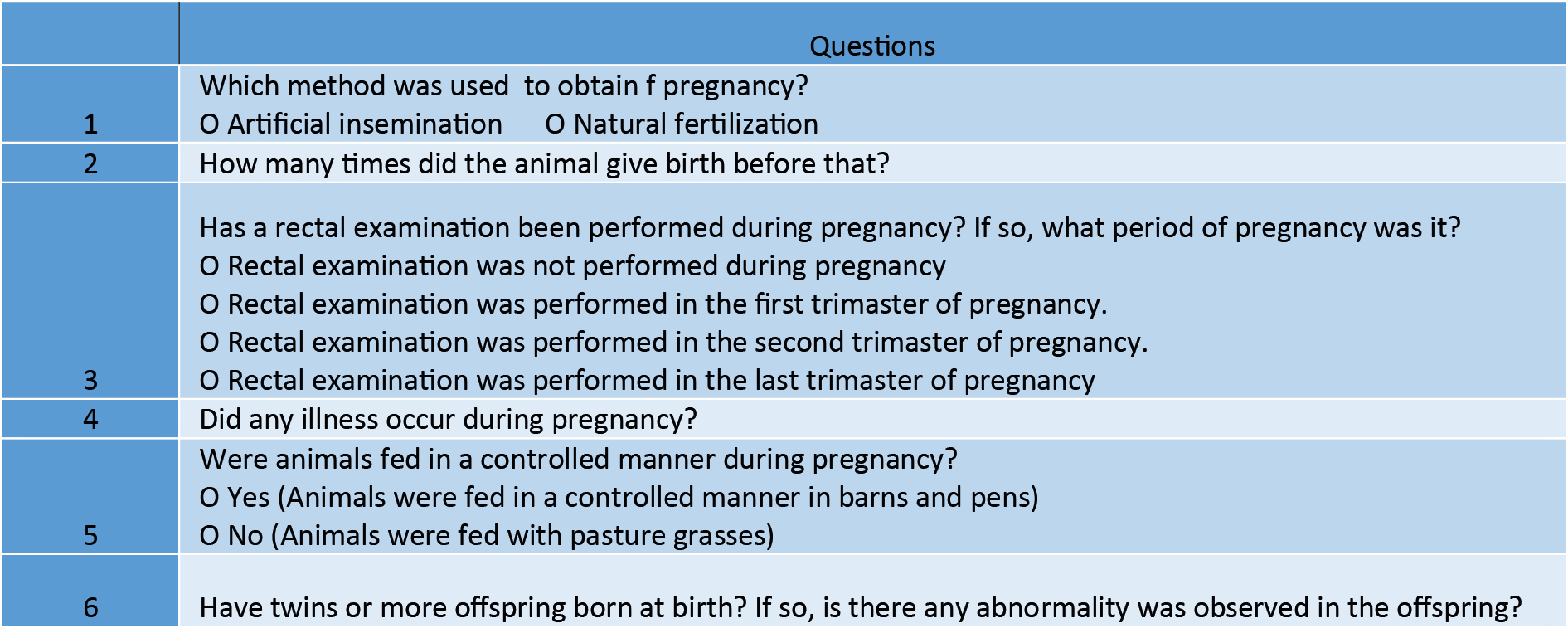
A total of 73 animals with congenital anomalies were examined macroscopically and an exahustive description of them were performed In each case, the specie, breed, sex, age and the origin were registered. In addition, a detailed and comprehensive questionary was performed in order to obtein more information for this study (Table 1).
The rates of congenital anomalies according to the species, breed, sex age and the organ/system affected were determined.
Results
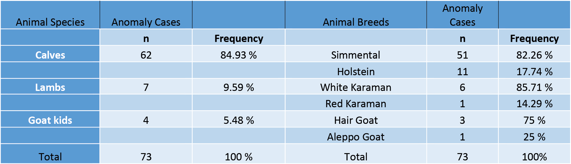
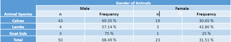
Between June 2020 and June 2021, descriptive study of congenital anomalies encountered in ruminant farms in Elazig region were performed. During the study, a total of 79 congenital anomalies were detected in 73 animals (Fig. 1- a,b,c,d). Congenital anomalies were detected in 84.93% (n= 62) calves, 9.59% (n= 7) in lambs, and 5.48% (n= 4) in goat kids. These anomalies were detected in 68.49% (n= 50) male animals and 31.51% (n= 23) in female animals. The distribution of congenital anomalies according to species, breed and gender are presented in table 2 and table 3.
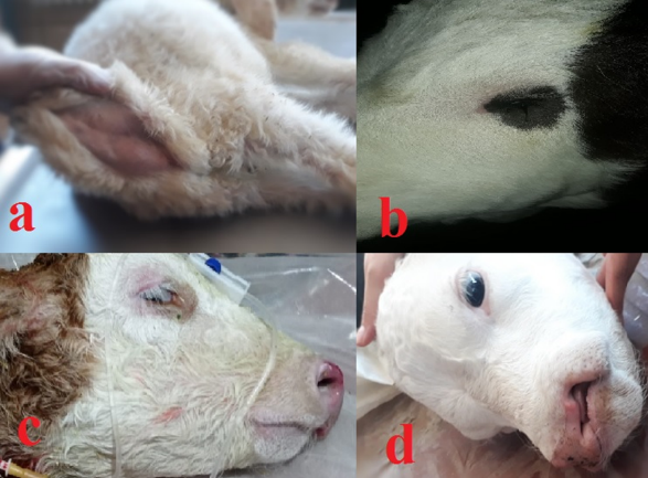
Figure 1. Some congenital anomalies: atresia ani (a) anophthalmia (b) mandibular malformation brachygnathi inferior (c) cheliognatopalatoschisis (d)
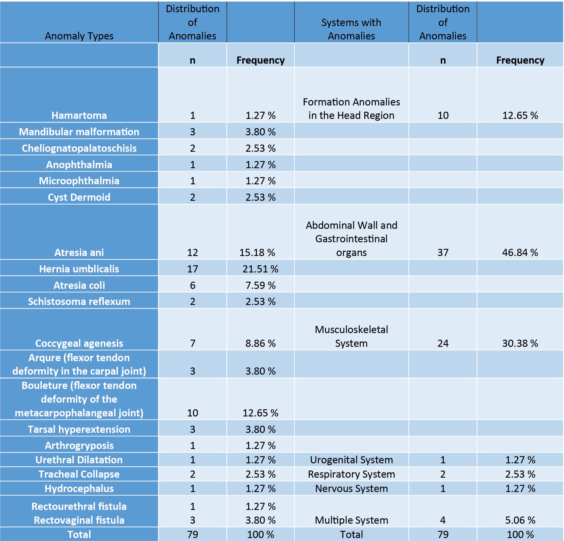
In this study, hernia umbilicalis (21.51%), atresia ani (15.18%) and bouleture (flexor tendon deformity of the metacarpophalangeal joint) (12.65%) were f the most common congenital anomalies found . The systems that most frequently included congenital anomalies were the abdominal wall-gastrointestinal system (46.84%) and the musculoskeletal system (30.38%). The distribution of congenital anomalies according to their types and systems involved is presented in Table 4.
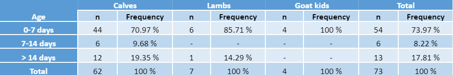
Most of congenital anomalies were detected between 0-7 days old (73.97%), and secondarily when the animals were more than 14 days old (17.81%).The relationship between the age and the presence of congenital anomalies in each animal species is presented in Table 5.
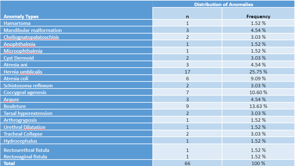
A total of 66 congenital anomalies were found in 62 calves. Most of them consisted of hernia umbilicalis (25.75%), bouleture (13.63%), cocygeal agenesis (10.60%) and atresia coli (9.09%). The distribution of congenital anomalies encountered in calves is presented in table 6.
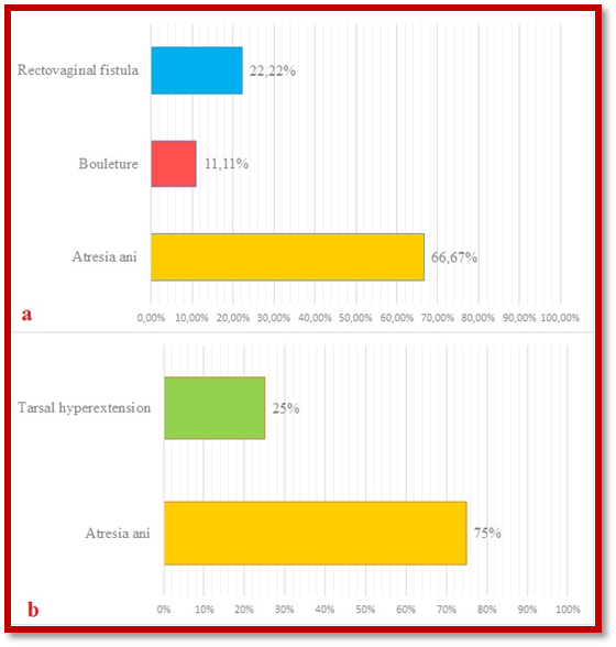
Atresia ani was the most common congenital anomaly in lambs (66.67%) and goat kids (75%). (Fig. 2- a,b).
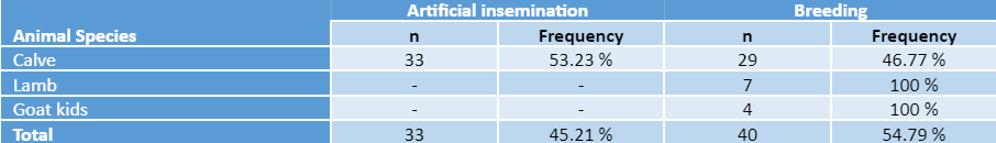
It was determined that 53.23% of cattle that had offspring with congenital anomalies were pregnant by artificial insemination. in contrast to sheep and goats who were pregnant by natural breeding. The relationship between the congenital anomalies of the offpringand the way of conception received in each animals species is presented in table 7.
Table 7. Relationship and distribution between the offspring with congenital anomalies and the way of conception in each animal species
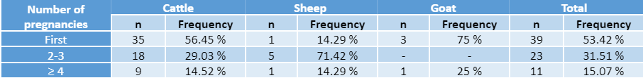
It was determined that the presence of offspring with congenital anomalies in cattle, sheep and goats in their first pregnancy was 56.45%, 14.29% and 75 %, in each case, respectively.. . It was found that 53.42% of all animals species had offspring with congenital anomalies in their first pregnancy . The relationship between the number of pregnancies and the offspring with congenital anomalies is presented in table 8.
Table 8. Relationship and frequency between number of pregnancies and offspring with congenital anomalies
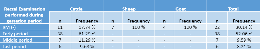
It was determined that 61.29% of cattle that had offspring with congenital anomalies received rectal examination for the diagnosis of pregnancy in the early period ofgestation . The relationship between the period of rectal examinations performed during pregnancy and the birth of offspring with congenital anomalies is presented in table 9.
Table 9. The relationship of rectal examination periods during pregnancy with birth of offspring with congenital anomalies
It was found that 83.56% of the animals that had offspring with congenital anomalies were grazing on pasture. It was determined that 82.19% of the animals did not have any disease during pregnancy.
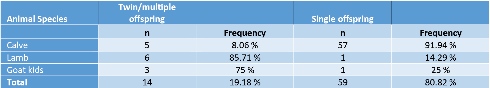
It was found that 19.18% of the animals with congenital anomalies were born as a result of twin (cattle) or multiple pregnancy (small ruminants). ,In small ruminants, the percentage of congenital anomalies was higher in lamb (85.71%) than goatkids (75%) (Table 10).
Discussion
Congenital anomalies are structural and functional pathologies related with genetic origin and/or environmental factors that acts during the pregnancy (Aksoy et al. 2006; Belge et al. 2000; Demiraslan et al. 2014; Kaya et al. 2011; Niwas et al. 2020; Tasal and Aytekin 2015). The incidence of congenital anomalies in ruminants ranges from 26.8% to 40.48% (Fubini 2004; Kaya et al. 2011; Ogurtan et al. 1997). Some congenital anomalies can cause death, while others can only create a structural defect without affecting vital functions (Kaya et al. 2011). In this study, the aime was to describe the main congenital anomalies encountered in ruminants from the Elazig region in Turkey.
Most of the congenital anomalies encountered in ruminants are seen in calves (Aksoy et al. 2006; Doğan and Sındak 2013). According to this, Aksoy et al. (2006) , reported that 83.3% of congenital anomalies encountered in ruminants in Kars region are seen in calves. In addition, this author (Aksoy et al. 2006), found that the rate of congenital anomalies was 14.8% in lambs and 1.9% in goat kids. Doğan and Sındak (2013), reported that 72.5% of congenital anomalies encountered in ruminants in Nizip region are seen in calves, being the rate of congenital anomalies of 7.5% in lambs and 20% in goat kids, respectively. In this study, it was found that 84.93% of congenital anomalies were seen in calves 9.59% in lambs and 5.48% in goat kids.The higher rate of congenital anomalies described by Doğan and Sındak (2013), in goats compared to other studies is related to the fact that the breeding of this species is higher in Nizip region compared to Elazig and Kars regions.
Some researchers reported that congenital anomalies encountered in calves are more common in Swiss Brown breed (Karabulut et al. 2001; Kaya et al. 2011). Some researchers reported that congenital anomalies are more common in Simmental calves.1 Kaya et al. (2011), reported that 47.42% of the congenital anomalies encountered in calves in Erzurum region occurred in the Swiss Brown breed. On the other hand, Karabulut et al.(2001), reported that 58.94% of the congenital anomalies encountered in calves in Elazig region occurred in the Swiss Brown breed. Aksoy et al. (2006), reported that 32.1% of congenital anomalies are in Simmental calves. Belge et al. (2000), reported that congenital anomalies are more common in crossbred calves. In this study, it was determined that 82.26% of the congenital anomalies encountered in calves occurred in the Simmental breed. All these studies suggest that congenital anomalies have not a breed predisposition. The reason for the difference between these studies is thought to be related with the populations of cattle breeds according to the regions.
Previous studies demostrated that the incidence of congenital anomalies is higher in male animals species (Aksoy et al. 2006; Belge et al. 2000; Kaya et al. 2011). Aksoy et al. (2006), in their study on ruminants in Kars region, reported that 67.55% of the animals with congenital anomalies were male, 64.67% were calves, 76.92% lambs, and 100% goat kids. Kaya et al. (2011), reported that 56.70% of the animals with congenital anomalies were male in their study on calves in Erzurum region. In a study conducted in the Van region, Belge et al. (2000), reported that 59.60% of the animals with congenital anomalies were males. . In this study, 69.35% of calves, 57.14% of lambs and 75% of goat kids were male.
Some researchers reported higher incidence of congenital anomalies in the musculoskeletal system (Kaya et al. 2011; Ogurtan et al. 1997; Ozaydın et al. 2006; Syed and Shanks 1992). In agreement with other authors (Aksoy et al. 2006, Dogan and Sındak 2013) in this studyt the prevalence of congenital anomalies in the abdominal wall and gastrointestinal system (46.84%) was higher than other types of anomalies. The rate of musculoskeletal anomalies was 30.38% being the second system with congenital anomalies.
It has been reported that the most common congenital anomalies in calves are hernia umblicalis, atresia ani and bouleture (Aksoy et al. 2006; Belge et al. 2000; Fubini 2004; Kaya et al. 2011; Lotfi and Shahryar 2009). However, Karabulut et al. (2001), reported that the most common anomalies in calves in Elazig region were bouleture (45.26%), cyst dermoid (17.89%) and arqure (17.89%). Doğan and Sındak (2013), reported that the most common congenital anomalies in calves in the Nizip region were hernia umblicalis and cyst dermoid. In this study, the most common were hernia umblicalis (25.76%), bouleture (13.63%) and cocygeal agenesis (10.60%). The rate of cyst dermoid cases was 3.03%.
Some researchers reported that the most common congenital anomalies in lambs are atresia ani, and in goat kids the urogenital system, respectively. (Aksoy et al. 2006; Doğan and Sındak 2013; Ogurtan et al. 1997). . In this study, it was determined that atresia ani was the most common cases of congenital anomaly in lambs (66.7%) and goat kids (75%) however, urogenital system anomalies were not observed in goat kids. .
In some studies (Aksoy et al. 2006; Bahr et al. 2003; Belge et al. 2000; Fırat et al. 2005; Harper et al. 1990; Miura et al. 1990; Ogurtan et al. 1997; Stanley 1993; Tsuda et al. 2004), it has been reported that the incidence of congenital anomalies is lower in the offspring of animals conceived by artificial insemination. In this study, it was determined that 53.23% of the calves with congenital anomalies became pregnant by artificial insemination being the incidence of congenital anomalies lower in offspring conceived by natural mating than in offspring conceived by artificial insemination.
Rectal examinations for pregnancy diagnosis performed in the early stages of gestation can be the cause of many congenital anomalies, especially intestinal atresia (Aksoy et al. 2006; Atalan et al. 2003; Belge et al. 2000; Demiraslan et al. 2014; Hendrickson et al. 1992; Kaya et al. 2011; Longeri et al. 2003; Newman et al. 1999). In this study, rectal examination was performed in the early period of gestation in 61.29% of cattle that had offspring with congenital anomalies, but atresi ani was not registered in these calves. Probably this practice was one of the predisposing factor for congenital malformations.
Congenital anomalies in calves and lambs were more frequently when they came from the first pregnancy of their dams, and in goat kids that they came from the second dam pregnancy. Most of small ruminant that had congenital anomalies came from dams with multiple gestation. frequently
Congenital anomalies are pathologies frequently present in ruminant breeding herds all over the world. Although the prevalence of congenital anomalies in ruminants varies according to the regions, it had been recorded between 26.8-40.48%. This study, describe the main congenital anomalies encountered in ruminants in the Elazig region of Turkey and the relationship between species, race, sex, age, number of pregnancies, rectal examination, and method of conception employed . More studies that include the participation of diagnostic laboratories are needed to improve the characterization of congenital anomalies that could contribute to the implementation of preventive and control measures

