Servicios Personalizados
Revista
Articulo
Indicadores
-
 Citado por SciELO
Links relacionados
-
 Similares en
SciELO
Compartir
Medicina (Buenos Aires)
versión impresa ISSN 0025-7680versión On-line ISSN 1669-9106
Medicina (B. Aires) v.69 n.5 Ciudad Autónoma de Buenos Aires set./oct. 2009
ARTÍCULO ORIGINAL
Transient expiratory flow interruptions (TEFI) increase expiratory flow in acute asthma exacerbation
Alejandra Fernández1, Sergio Guardia1, Alejandra González1, Roberto Serrano1, Daniel Rodenstein2, Hernando Sala1
1Servicio de Neumonología, Hospital Posadas, El Palomar, Buenos Aires;
2Pneumonology Unit, Saint Luc Hospital, Catholic University of Louvain, Brussels, Belgium
Dirección postal: Dr. Hernando Sala, Delgado 925,1428 Buenos Aires, Argentina, Fax: (54-11) 4469-9202 e-mail: nardosala@gmail.com
Abstract
We have shown that expiratory flows increase when expirations are rapidly interrupted in stable asthmatic patients. We hypothesized that a similar increase could be obtained in patients with acute exacerbation of bronchial asthma treated in the Emergency Room. A total of 30 asthmatic patients were randomly allocated into two groups, the study and the control groups. Patients in the study group were connected to a device with an inspiratory line designed to administer pressurized aerosols. The expiratory line passed through a valve completely interrupting flow at 4 Hz, with an open/closed time ratio of 10/3. The control group patients were also connected to the device, but with the valve kept open. Mean expiratory flow at tidal volume (MEFTV) was measured under basal conditions and at 4, 8 and 12 minutes after connecting the patients to the device. All patients received standard treatment throughout the procedure. At all time points MEFTV increased more in the study than in the control group (p < 0.003 by two-way ANOVA). There was no residual effect after disconnection from the device. We conclude that TEFI can rapidly improve expiratory flows in patients with acute exacerbations of asthma, while pharmacologic interventions proceed.
Key words: Asthma; Expiratory flow; Lung heterogeneity; Pendelluft
Resumen
Incremento del flujo espiratorio luego de interrupciones transitorias en la exacerbación aguda de asma. Demostramos que el flujo espiratorio máximo, en pacientes asmáticos en estado estable, se incrementaba cuando se generaban rápidas y transitorias interrupciones del flujo. Formulamos la hipótesis de que un incremento similar podría ser observado en pacientes con exacerbación aguda de asma tratados en la sala de emergencias. Un total de 30 pacientes asmáticos fueron distribuidos al azar en dos grupos. Los pacientes del grupo en estudio fueron conectados a un aparato con una vía inspiratoria diseñada para la administración de aerosoles. La vía espiratoria pasaba por una válvula que interrumpía el flujo completamente a 4 hz, con una relación tiempo abierta/tiempo cerrada de 10/3. Los pacientes del grupo control también fueron conectados al aparato pero con la válvula siempre abierta. Se midió el flujo medio de la espiración a volumen circulante en condiciones basales y a los 4, 8 y 12 minutos después de conectado el paciente al equipo. Todos los pacientes recibieron el tratamiento farmacológico estándar a lo largo del ensayo. Se observó un incremento significativamente mayor del flujo espiratorio medio a volumen circulante en el grupo en estudio en comparación al grupo control (p < 0.003 ANOVA de dos vías) durante todo el ensayo. No hubo efecto residual después de la desconexión del equipo. Concluimos que las interrupciones transitorias del flujo espiratorio pueden incrementar rápidamente el flujo espiratorio en pacientes con exacerbaciones agudas de asma dando tiempo a que el tratamiento farmacológico comience a actuar.
Palabras clave: Asma; Flujo espiratorio; Heterogeneidad pulmonar; Pendelluft
Despite the recent advances in the pharmacological treatment of bronchial asthma, the incidence of the disease still increases and acute exacerbations are still the hallmark of the disease. Severe acute asthma is treated with beta mimetics, inhaled and oral corticosteroids, and other pharmacological means1. It might be possible also to alleviate flow limitation by using mechanical means2-4. The main observation that let us to our present hypothesis was that in stable state asthmatic patients the measured FEV1 increased when maximal expiratory efforts were performed with rapid brief repeated interruptions of expiration, in comparison with basal non-interrupted efforts5.
Since during acute exacerbations of bronchial asthma flow limitation is so great that flow may be limited even during tidal breathing, it may well be that expiratory flow could increase if tidal expiration was rapidly interrupted with an electromechanical valve. To include patients with severe enough flow limitation we chose to study patients admitted, for acute exacerbation of bronchial asthma, to the emergency room.
Materials and Methods
Thirty consecutive patients attending the emergency room on two predetermined days, when the investigation team was available, from 9 am to 3 pm were included. They met inclusion and exclusion criteria and accepted to participate in the study after signing an informed consent form.
The patients had a clear history of asthma exacerbation with dyspnea and wheezing, obstructive pattern on spirometry including an FEV1 < 80% of reference values, with no history of tobacco smoking or other pulmonary or cardiac diseases that could be considered confounding factors. These patients were randomly allocated into two groups with 15 patients each, the study group and the control group. The protocol had been approved by the Posadas Hospital Human Ethics Committe.
Clinical examination and spirometry were performed at entry into the study. Transcutaneous O2 saturation was measured throughout the study (Criticare 504 Systems, inc.). Baseline spirometry was performed according to the ATS criteria6 by means of a rolling seal spirometer with a flat frecuency response up to 7 Hz, a resistance of less than 2 cm H2O.L.s-1 and a flow of 12 L.s-1 (Sensormedics, USA), predicted spyrometric values were calculated using the 1993 European Respiratory Society regression equations. Immediately after, methylprednisone 40 mg. was orally administrated to the patients, who were connected to a device (Fig. 1) consisting of a 4 cm internal diameter inspiratory line, suitable for pressurized aerosols delivery and oxygen administration. The inspiratory line was connected to patients by a low resistance unidirectional 3-way valve (Hans Rudolph, USA). The expiratory line passed through a specially designed electromechanical valve. The valve consisted in a central axis with two wings with alternating swinging movement that opened and successively closed the expiratory line. The movement was regulated by a potentiometer which was controlled by an electronic circuit. The latter defined the cycling frequency and the percentage of time the valve remained closed. In this experiment it was set at a cycling frequency of 4 Hz and an open/closed time ratio of 10/3. Indeed, complete valve opening and closing times were 16 miliseconds (ms), 4 interruptions taking place in one second, the closed time was 59 ms, the open time was 159 ms. Therefore, 23.6% of each cycle the valve was closed, 63.6% the time the valve was opened and 12.8% of the time the valve was opening or closing. Beyond the valve, the expiratory line was connected to a manually operated three-way valve connected to either the rolling seal spirometer or to room air.

Fig. 1.- Schematic diagram of the TEFI device. A: Inspiratory line, B: Pressurized aerosol. C: Unidirectional 3-ways valve. D: Mouthpiece. E: Expiratory line. F: Electromechannical valve. G: Manually activated 3-ways valve. H: Rolling-seal spirometer I: Room air
During tidal breathing, the expiratory volume was continuosly measured and divided by the expiratory time to yield a "mean expiratory flow at tidal volume (MEFTV)". In addition, expiratory flow-volume curves are constructed from volume and the derived flow. This was recorded during 3 consecutive respirations in both groups, with the valve left open, and then, the expiratory line was connected to room air. In the control group the valve was left open. In the study group the cycling valve was activated. The MEFTV measurements were repeated 4, 8 and 12 minutes after the initial measurement. After 13 minutes, patients were disconnected from the device. The FEV1 determinations were repeated 20 minutes from the start of the procedure.
In the first 13 patients included in the protocol (6 from the control group and 7 from the study group), when baseline spirometry was performed the tidal breathing flow-volume curve was recorded immediately before the maximal flow-volume curve. The expiratory flow of both curves, for the same lung volume, were visually compared.
Repetitive administration of salbutamol (total dose 800 mcg), ipratropium bromide (total dose 168 mcg), and budesonide (total dose 1600 mcg) were performed through pressurised cannisters (4 puffs at minute 0, 2 puffs at minute 4 and 2 puffs at minute 12 of each drug).
Statistical analysis was performed with SPSS. Comparisons between baseline and final FEV1 was determined by paired Student t test. Comparisons between groups were performed with unpaired Student t test. MEFTV logarithmic transformation was performed in order to normalize the distribution of data and allow parametric analysis application by means of a two-way ANOVA test.
Results
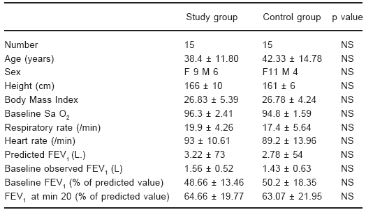
Average anthropometric, clinical and spirometric data of both groups are shown in Table 1. No differences between groups were found in age, sex distribution, baseline O2 saturation, FEV1 in percent of predicted value, respiratory or cardiac rates. Predicted FEV1 in absolute value of the control group was slightly lower but not significantly so (p < 0.07).
TABLE 1.- Anthropometric clinical and spirometric data of both groups

Data are presented as mean ± SD; p values < 0.05 were considered statistically significant.
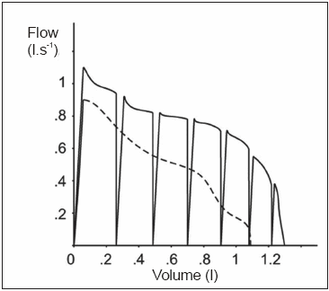
The entire population showed significant increases in FEV1 (mean ± SD) at 20 minutes (31.24 ± 23%) in comparison with baseline FEV1 (p < 0.001) but no difference between groups was observed (31.87 ± 16% for the study group vs. 30.57 ± 29% for the control group). Figure 2 shows the comparison of basal tidal expiratory flow vs. expiratory flow at minute 4 in the patient who improved the most when interruptios of expiratory flow was performed.

Fig. 2.- Expiratory tidal flow - volume curve at minute 0 (dotted line) and at minute 4 (continuous line) in the patient who improved the most in the treatment group.
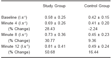
The mean expiratory flow at tidal volume was slightly but not significantly higher at baseline in the study group, and increased significantly more in the study group than in the control group (p < 0.003 two-ways ANOVA. Table 2 and Fig. 3). In fact, the control group showed a maximal increase in MEFTV of 16.44% (at 12 minutes), whereas the study group showed a maximal increase of 50.68% at the same time measurement (Fig. 3). No differences between groups were observed in tidal volume across the study (two ways ANOVA).
TABLE 2.- Change in mean expiratory flow at tidal volume in both groups

Data are presented as mean ± SD in abolute value and percentage of change. Comparison between groups was performed by two ways ANOVA (p < 0.003)

Fig. 3.- Comparison between groups in the change in % of MEFTV ± SEM over time. Study group (![]() ) Control group (
) Control group (![]() ) (p < 0.003 two-ways ANOVA).
) (p < 0.003 two-ways ANOVA).
No correlation was found between the baseline FEV1 in percentage of predicted values and the change in mean MEFTV.
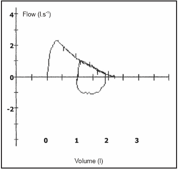
Figure 4 shows the tidal and maximal flow-volume curves at baseline in one of the 13 subjets where this was measured. As it can be seen, tidal expiratory flows were maximal, i.e. coincided with the flows obtained during a maximal expiration. The first 13 patients included in the study did not differ in baseline FEV1, FEV1% predicted, respiratory and cardiac rates, or any others anthropometrics index from the latests 17 patients whom only maximal flow-volume curve were recorded.

Fig. 4.- Tidal expiratory flow - volume loop and maximal expiratory curve preformed immediately after in one of the 12 out of 13 patients in which overlaps of both curves were observed.
There were no adverse or deleterious effects related with the TEFI technique.
Discussion
We have found that rapid repetitive interruptions of expiratory flow during tidal breathing in patients with acute exacerbations of bronchial asthma allow for improvements in expiratory flows. The improvement is immediate but there is no residual effect once the patients are disconnected from the device. The better result in MEFTV in the study group was obtained despite the fact that the baseline value was already higher in this group.
Expiratory airflow limitation is one of the main physiologic characteristic of asthma exacerbation. It is also probably one of the main determinant of dyspnea, the most disabling symptom of the disease. Experience with non-pharmacologic interventions in asthma exacerbations is not large and included continuous positive airways pressure4 and bilevel noninvasive positive pressure ventilation3. The technique here presented, TEFI, appears as capable of rapidly increasing expiratory flow at tidal volume respiration. Even if the effect vanishes upon disconnection from the device, TEFI may allow to "buy time" while pharmacologic therapy produces a more fundamental improvement in the status of the patient.
Before discussing the results some methodological aspects must be taken into account. We used a rolling seal spirometer, which inertial and response time characteristics seem adequate for the dynamic constraints of our valve. In addition, it has better noise/signal ratio than the pneumotachographs when the valve is cycling7. Mean expiratory flow at tidal volume seems to be a useful parameter to continuously monitor flow limitation over time in asthmatic patients, at least from a pragmatic point of view. It obviates the need for forced inspirations and expirations for measurements of FEV1, that can be difficult to perform for asthmatic patients in acute exacerbation and that can even worsen bronchospasm in some patients8-10. In order to demonstrate that asthmatic patients, during severe acute exacerbations, like the one observed in the patients that we have studied, have expiratory flow limitation at tidal breathing, we compared expiratory flows during tidal and maximal expirations at the same lung volume (Fig.5). We found that in12 out of 13 subjects where this comparison was made, tidal expiratory flows were identical to the maximal flow at the corresponding volume of the maximal expiratory flow- volume curve11.
Other methods like airway resistance measurement by forced oscillation technique cannot be used while the valve is cycling at 4 Hz, because of the noise generated by the very similar FOT device loudspeaker cycling frequency12. The mechanical valve does not interfere with the inspiratory line, therefore deposition of drugs given by aerosols through the inspiratory line are not altered.
Present results extend previous ones demonstrated in asthmatic patients in stable state and in normal subjects5, 7. The phenomenon of supramaximal flows has been studied in the last century both during partial maximal flow-volume curves and during natural coughing. It designates expiratory flows that exceed maximal expiratory flows, which are deemed to represent the mechanical characteristics of the airways under conditions of maximal expiratory effort. Partly, supramaximal flows are due to the compression of the so called flow limiting segment, located generally in the large central airways. Asthmatic patients increased resistance induces choke point upstream migration and airways collapse even at tidal volume13. This situation changes during flow interruption and airways geometry goes back to no flow conditions. When flow restarts the airways again collapse and the air included in the flow limiting segment contributes to supramaximal flow as Knudson et al. have demonstrated14. However, Pedersen et al. found that the air volume included in supramaximal flows was greater than the air contained in the flow limiting segment15. So, they suggested that during the transition from an unstable configuration to a stable collapsed configuration the airway morphology permits higher flows. This mechanism is probably important in the response of asthmatic patients to TEFI.
Studying normal subjects and patients with airway obstruction, the response to interrupted and non interrupted expiratory flow-volume curves, from total lung capacity to residual volume5, 7 we observed a significant correlation between interruption time and supramaximal flows measured immediately after. The larger the interruption time, the higher supramaximal flows. This was also the case in a lung mechanical model16. We have also shown that in normal subjects and in asthmatic patients supramaximal flows correlate with flow limitation, supramaximal flows increase as FEV1 and FEF 25-75, in % of reference value, decrease. This correlation was not found in the present study but this time we have not measured maximal forced expirations.
We propose that during the interruption time, air moves from the less emptied slow lung units, with longer time constant, to the more emptied fast lung units, a mechanism called "pendelluft", consequence of the smaller lung recoil pressure of the fast units with small relative volume in comparison with slow lung units. When flow restarts the fast units can contribute more to total flow, thus increasing supramaximal flows7, 17, 18. Mead and Whittenberger have shown that approximately 18 msc are necessary to equilibrate alveoli- mouth pressures during a total occlusion at the mouth. This figure includes lung tissue resistance and airway resistance19. Our interruptions, lasting 59 msc, could therefore give enough time for equilibration within the lungs following the «pendelluft» mechanism.
It is probable that other mechanisms can play a role in response to TEFI. Airway resistance was not measured and so changes in smooth muscle tone, oedema and secretions were not assessed.
Very recently Downie et al.20 demonstrated that baseline ventilation heterogeneity is a strong predictor of airway hyperresponsiveness, a more uneven distribution of ventilation and airway narrowing throughout the lung results in a greater overall increase in airway resistance, and they suggested that normalisation of ventilation heterogeneity is therefore a potential goal of treatment. In our opinion, pendelluft during airflow interruption can partially compensate for the heterogeneity of the asthmatic lung by allowing slow units to "use" the better mechanical characteristics of fast units to improve lung emptying.
In asthma exacerbation the decrease in flow limitation could lead to a decrease in the respiratory load and so diminish the risk of respiratory failure due to muscle fatigue.
Nevertheless, even if the physiological effect of TEFI appears as beneficial, the potential clinical improvement still needs to be ascertained.
Acknowledgements: The authors thank F. Rios and Prof. G. Liistro for statistical analysis, J.Segovia for his cooperation in patients recruitment, and G. Guardia and P. Luque for technical support.
Conflicts of interest: None declared.
1. Global Strategy for asthma management and prevention. Rev 2006; cap 3, 27-40. [ Links ]
2. Qvist J, Andersen J B, Pemberton M, et al. High-level PEEP in severe asthma. N. Engl J Med 1982; 307: 1347-8. [ Links ]
3. Soroksky A, Stav D, Shpirer I. A pilot prospective, randomized, placebo-controlled trial of bilevel positive airway pressure in acute asthmatic attack. Chest 2003; 123: 1018-25. [ Links ]
4. Rodrigo G, Rodrigo C, Hall JB. Acute asthma in adults: A Review. Chest 2004; 125: 1081-1102. [ Links ]
5. Sala H, Fernandez A, Guardia S, et al. Supramaximal flow in asthmatic patients. Eur Respir J, 2002; 19: 1003-7. [ Links ]
6. ATS. Standarization of spirometry. 1994 Update. Am J Respir Crit Care Med 1995; 152: 1107-36. [ Links ]
7. Sala H, Galindez F, Badolati A, et al. Relationship between supramaximal flows and flow-limiting mechanisms. Eur Respir J 1996: 9, 512-6. [ Links ]
8. Beaupré A, Orehek J. Factors influencing the bronchodilator effect of a deep inspiration in asthmatic patients with provoked bronchoconstriction. Thorax 1982; 37: 124-8. [ Links ]
9. Lim TK, Pride NB, Ingram RH. Effects of volume history during spontaneous and acutely induced airflow obstruction in asthma. Am Rev Respir Dis 1987; 135: 591-6. [ Links ]
10. Lim TK, Ang SM, Rossing TH, et al. The effects of deep inhalation on maximal expiratory flow during intensive treatment of spontaneous asthmatic episodes. Am Rev Respir Dis 1989; 140: 340-3. [ Links ]
11. Hyatt RE. Expiratory flow limitation. J Appl Physiol.1983; 55: 1-8. [ Links ]
12. Navajas D, Farré R. Oscillation mechanics. In: Milic Emili J, (ed.) European Respiratory Society Journals Ltd., 1999, pp 112-3. [ Links ]
13. Dawson S.V. Elliot E.A. Wave-speed limitation on expiratory flow-a unifying concept. J Appl Physiol 1997; 43: 498-515. [ Links ]
14. Knudson RJ, Mead J, Knudson DE. Contribution of airway collapse to supramaximal expiratory flows. J Appl Physiol 1974; 36: 653-67. [ Links ]
15. Pedersen OF, Lyager S, Ingram RH Jr. Airway dynamics in transition between peak and maximal expiratory flow. J Appl Physiol 1985; 59: 1733-45. [ Links ]
16. Sala H, Veritier C, Rodenstein D, et al. Can «pendelluft» explain supramaximal flows during interrupted forced expirations. Eur Respir J Suppl 1999; 14: Suppl. 30, 393. [ Links ]
17. Sala H, Gonzalez A, Fernandez A, et al. Supramaximal flow after salbutamol and methacholine in asthmatic patients. Am J Respir Crit Care Med 1998; 157: A88. [ Links ]
18. Gonzalez A, Fernandez A, Barañano C, et al. Supramaximal flow: Comparison between asthmatic and COPD patients. Eur Respir J Suppl 1999; 14: Suppl. 30, 81. [ Links ]
19. Mead J, Whittenberger JL. Evaluation of airway interruption technique as a method for measuring pulmonary air-flow resistance. J Appl Physiol 1954; 7: 408-16. [ Links ]
20. Downie SR, Salome CM, Verbanck S, et al. Ventilation heterogeneity is a major determinant of airway hyperresponsiveness in asthma, independent of airway inflammation.Thorax 2007; 62: 684-9. [ Links ]
Recibido: 8-7-2008
Aceptado: 2-12-2008


