Servicios Personalizados
Revista
Articulo
Indicadores
-
 Citado por SciELO
Links relacionados
-
 Similares en
SciELO
Compartir
Archivos argentinos de pediatría
versión impresa ISSN 0325-0075
Arch. argent. pediatr. vol.111 no.5 Buenos Aires oct. 2013
http://dx.doi.org/10.5546/aap.2013.418
ORIGINAL ARTICLE
http://dx.doi.org/10.5546/aap.2013.418
Fetal and neonatal mortality in patients with isolated congenital heart diseases and heart conditions associated with extracardiac abnormalities
Pablo Marantz, M.D.a, M. Mercedes Sáenz Tejeira, M.D.a, Gabriela Peña, M.D.a, Alejandra Segovia, M.D.a, and Carlos Fustiñana, M.D.b
a. Pediatric cardiology department.
b. Neonatology Division.
Hospital ltaliano de Buenos Aires, Buenos Aires, Argentine.
E-mail Address: Pablo Marantz, M.D.: pablo.marantz@hospitalitaliano.org.ar
Conflict of Interest: None.
Received: 01-19-2012
Accepted: 05-03-2013
ABSTRACT
Congenital malformations are a known cause of intrauterine death; of them, congenital heart diseases (CHDs) are accountable for the highest fetal and neonatal mortality rates. They are strongly associated with other extracardiac malformations and an early fetal mortality. Two hundred and twenty fves cases of CHDs are presented. Of them, 155 were isolated CHDs (group A) and 70 were associated with extracardiac malformations, chromosomal disorders, or genetic syndromes (group B). The overall mortality in group B was higher than that observed in group A (p <0.01). Prenatal mortality was similar in both groups: A: 8.4% (13 out of 155); B: 15.7% (11 out of 70). Postnatal mortality was A: 16.8% (26 out of 155) (p <0.01), OR: 0.52 (95% CI: 0.16-1.7); B: 32.9% (23 out of 70) (p <0.01), OR: 0.41 (95% CI: 0.20-0.83). Heart diseases associated with extracardiac abnormalities had a higher mortality rate than isolated congenital heart diseases in the period up to 60 weeks of postmenstrual age (140 days post-term). No differences were observed between both groups of patients in terms of prenatal mortality.
Key words: Survival; Prenatal diagnosis; Fetal mortality; Congenital heart diseases.
INTRODUCTION
Congenital malformations are a known cause of intrauterine death.1 Of them, congenital heart diseases (CHDs) are accountable for most cases of fetal and neonatal mortality.2
CHDs are greatly associated with other extracardiac malformations and, according to published data, they lead to a high fetal mortality rate;5-8 for this reason, we were interested in studying whether the presence of these findings modified prenatal and postnatal development.
The study objective was to assess prenatal and postnatal mortality caused by isolated heart diseases or when associated with other extracardiac malformations, from prenatal diagnosis up to 60 weeks of postmenstrual age (140 days post-term).
MATERIAL AND METHODS
This was an observational, prospective, cohort study. Between March 2003 and July 2009, all fetuses of pregnant patients aged 18-44 years old, referred to the Fetal Medicine Unit (FMU) of Hospital ltaliano de Buenos Aires, at 18-38 weeks of gestation were examined. Subjects included were pregnant women with a suspected alteration in the four-chamber and three-vessel view of the fetal heart during the routine fetal ultrasound examination, or with a history of CHD risk (pregnant women with a metabolic condition, severe obesity, connective tissue disease, exposure to a teratogenic agent, suspected chromosomal abnormality or extracardiac malformation, family history of CHDs, fetal infection, oligohydramnios-polihydramnios, and multiple pregnancy). A pediatric cardiologist performed a Doppler echocardiogram on the fetuses of these patients in order to confirm or rule out the presence of a heart condition. A real time General Electric Vingmed Vivid V ultrasound device with 1.5-3.6 mHz and 4.4-10 mHz cardiac transducers was used.
Pregnant women at risk of being lost to follow-up during the study and those with an inadequate ultrasound window during obstetric or fetal cardiac ultrasounds were excluded.
The suspicion of a genetic syndrome was confirmed or ruled out during the neonatal period by the hospital's Genetic Medicine Department, or postmortem by the Anatomic Pathology Service.
In order to calculate the sample size, a mortality difference over 20% with a 95% confidence interval and an 80% power was estimated, with a minimum of 70 patients per group.
The sample was divided into two groups: group A, patients with isolated CHDs; group B, patients with CHDs associated with other extracardiac malformations, chromosomal abnormalities or genetic syndromes. Survival and mortality were analyzed in the period between the prenatal diagnosis and up to 60 weeks of postmenstrual age (140 days post-term) or the time of death.
Data were analyzed using the c2 test for dichotomous outcome measures, and expressed using odds ratios with 95% confidence intervals.
Survival over time was analyzed using the Kaplan-Meier test.
RESULTS
A total of 1658 fetuses were examined at the FMU using a fetal echocardiogram; CHDs were detected in 225 of them.
Groups were as follows: group A, patients with isolated CHDs (n= 155); group B, patients with CHDs associated with other extracardiac malformations, chromosomal abnormalities or genetic syndromes (n= 70). Overall mortality in group B was higher, with a significant difference (p< 0.01) from group A (Table 1). When prenatal mortality was assessed, it was similar between both groups (Table 1): 8.4% (13 out of 155) in group A and 15.7% (11 out of 70) in group B. Postnatal mortality was 16.8% (26 out of 155) in group A (p <0.01), OR: 0.52 (95% CI: 0.16-1.7) and 32.9% (23 out of 70) in group B (p <0.01), OR: 0.41 (95% CI: 0.20-0.83).
Table 1. Overall prenatal and postnatal mortality in groups A and B

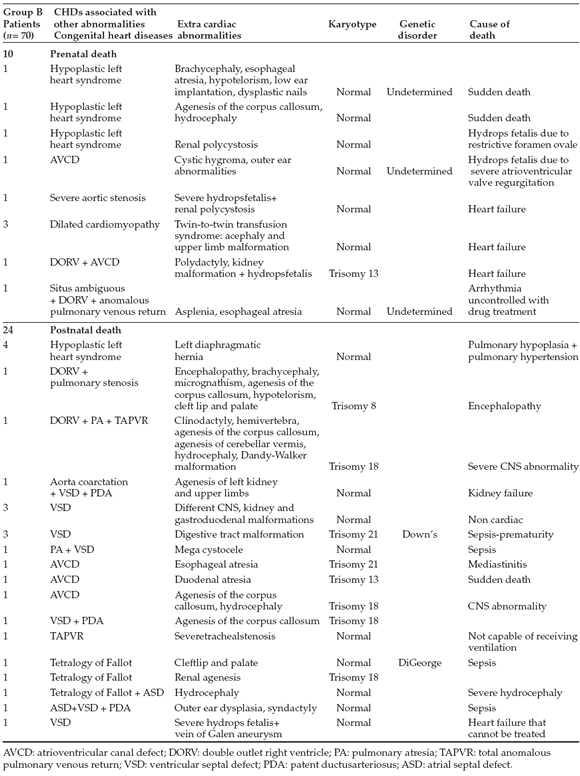
Tables 2 and 3 describe CHDs found in the deceased fetuses and newborn infants of groups A and B, respectively, and their extracardiac
Table 2. Isolated congenital heart diseases. Mortality

Table 3. Congenital heart diseases associated with extracardiac malformations, chromosomal abnormalities or genetic syndromes in deceased patients from group B

The analysis of postnatal mortality in group A indicated that 26 newborn infants died; 10 out of 155 (6.5%) during the postoperative period, and 16 out of 155 (10%) who had not undergone a heart surgery either due to their severity or because of the absence of a surgical indication. These data and the causes of death are detailed in Table 2: 8 patients had hypoplastic left heart syndrome (HLHS) and could not undergo surgery due to their critical status; 4 patients had severe Ebstein's anomaly and died due to heart failure and pulmonary hypertension with no response to medical treatment, 2 of them were preterm newborn infants. One patient with tetralogy of Fallot and pulmonary valve agenesis died due to sepsis.
This series presents the association of different chromosomal abnormalities and congenital heart diseases, which are detailed in Table 4. In group B, an association was found with an abnormal kariotype in 11 out of 34 cases (32.3%) and with non-chromosomal genetic syndromes in 4 out of 34 cases (8.8%). Chromosomal abnormalities were trisomy 18 and 21, with 4 cases each. In addition, 1 trisomy 8 case and 2 trisomy 13 cases were found. In relation to non-chromosomal genetic syndromes, there were 4 cases presented: in 1 of the cases DiGeorge syndrome was diagnosed, and 3 could not be included in any known syndrome.
Table 4. Congenital heart diseases associated with chromosomal disorders

Extracardiac abnormalities were related to the central nervous system (CNS) in 8 out of 34 cases (23%), followed by digestive tract malformations in 5 out of 34 cases.
DISCUSSION
In this cohort, the survival rate of patients with isolated CHDs was significantly higher than that of patients with CHDs associated with an extracardiac abnormality: 74.8% (116 out of 155) versus 51.4% (36 out of 70).
The association with extracardiac abnorma-lities was one of the factors with an impact on mortality, either due to the overall severity of the patient's condition or because a long-term prognosis was contradictory to invasive treatment. There were no significant differences in terms of CHD type, frequency and severity; therefore, we assumed that group B was made up of CHDs that should have similar results to group A in relation to their survival.
Although the association with extracardiac malformations casts a shadow over the prognosis, it is known that this is not the only outcome measure to be taken into consideration. It should be noted that prematurity and low birth weight were not analyzed in this study, and it is believed that they could have had a significant influence on morbidity and mortality, as well as on the final outcome.
In this series, no significant differences were observed in prenatal mortality (24.1%), including both groups. The rate was lower than that described by some large European studies,5,9,10 where pregnancy termination is a common practice, unlike the situation in Argentine.
It is interesting to note that in group B, 10 out of 70 fetuses died in the prenatal stage; all of them had a normal karyotype, but 3 were carriers of an unidentified genetic syndrome, and their death was because of heart failure. One case was associated with a situs ambiguous and an arrhythmia that could not be treated pharmacologically; the other case was an atrioventricular canal defect with severe regurgitation of the common AV valve.
The rate of postnatal mortality in group B (48.6%) was similar to that observed in the studies made by Eronen1and Boldt,3 who described a mortality rate close to 44%. These authors attribute this high mortality rate to the complexity of the heart disease, but also to its association with extracardiac and chromosomal malformations.
As a result, we believe that pregnant women and their unborn children should be timely referred to a facility with a higher level of care where they can be managed by a multidisciplinary team of professionals, taking into account that the best incubator for such babies is their mother's womb.
When analyzing extracardiac abnormalities frequency and characteristics, the most common were chromosomal abnormalities, followed by multiple extracardiac malformations and, to a lesser extent, non-chromosomal genetic syndromes. The incidence of chromosomal abnormalities among the deceased cases in group B was 29%; in other studies, such incidence ranged between 28% and 66%.3-6,12-15
We agree with most authors that CNS abnormalities are the ones most commonly associated with CHDs; in our series, the agenesis of the corpus callosum was the most common abnormality, but other studies have found that anencephaly, hydrocephaly,and spina bifida were the most common findings.8,15
CONCLUSIONS
In this study, heart diseases associated with extracardiac abnormalities had a higher morbidity and mortality rate than isolated congenital heart diseases in the period up to 60 weeks of postmenstrual age (140 days post-term).No differences were observed in prenatal mortality when comparing both groups of patients.
Acknowledgments
To Marianna Guerchicoff, M.D., for collaborating in data collection and manuscript editing.
1. Alfredo Ovalle S, Elena Kakarieka W. Estudio anátomo-clínico de las causas de muerte fetal. Rev Chil Obstet Gine-col 2005;70(5):303-12. [ Links ]
2. Guerchicoff M, Marantz P. Evaluación del impacto del diagnóstico precoz de las cardiopatías congénitas. Arch Ar-gent Pediatr 2004;102(6):445-50. [ Links ]
3. Boldt T, Andersson S, Eronen M. Out come of structur-al heart disease diagnosed in utero. Scand Cardio vasc J 2002;36(2):73-9. [ Links ]
4. Copel JA, Tan AS, Kleinman CS. Does a prenatal diagnosis of congenital heart disease altershort-termoutcome? Ultrasound Obstet Gynecol 1997;10:237-41.
5. Tennstedt C, Chaoui R, Korner H, Dietel M. Spectrum of congenital heart defects and extracardiac malformations associated with chromosomal abnormalities: result of a sevenyearnecropsystudy. Heart 1999;82:34-9.
6. DeVore GR.The role of fetal echocardiography in genetic sonography. Semin Perinatol 2003;27(2):160-72. Review.
7. Yagel S, Cohen SM, MessingB. First and early second tri-mester fetal heart screening. Curr Opin Obstet Gynecol 2007;19(2):183-90.
8. Lombardi CM, Bellotti M, Fesslova V, Cappellini A. Fetal echocardiography at the time of the nuchal translucency scan. Ultrasound Obstet Gynecol 2007;29(3):249-57. [ Links ]
9. O'Brien SM, Jacobs JP, Clarke DR, Maruszewski B, Jacobs ML, etal. Accuracy of the aristotlebasic complexity score or classifying the mortality and morbidity poten-tialof congenital heart surgery operations. Ann ThoracSurg 2007;84(6):2027-37.
10. Garne E and the Eurocat Working Group. Prenatal diagnosis of six major cardiac malformations in Europe - A popula-tion- based study. Acta Obstet Gynecol Scand 2001;80:224-8. [ Links ]
11. EronenM,etal.Outcomeoffetuseswithheartdisease-diagnosed in utero. Arch Dis Child Fetal Neonatal Ed 1997;77(1):F41-6.
12. Departament of Patholgy, Charite Hospital of the Univer-sity, Berlin, Germany. Spectrum of congenital heart defects ad extra cardiac malformations associated with chromo-somal abnormalities: results of a seven years necropsy study. Heart 1999;82(1):34-9. [ Links ]
13. Chaoui R, Korner H,Bommer C, Goldner B, et al. Heart de-fects and associated chromosoma laberrations. Ultraschall Med 1999;20(5):177-84. [ Links ]
14. Pepes S, Zidere V, Allan L D. Prenatal diagnosis of left atrial isomerism. Heart 2009;95(24):1974-7. Epub.
15. Tegnander E, Williams W, Johansen O J, Blaas H G, Eik-Nes SH. Prenatal detection of heart defects in a non-selected population of 30, 149 fetuses-detection rates and out come. Ultrasound Obstet Gynecol 2006;27(3):252-65. [ Links ]


