


Services on Demand
Journal

Article
 English (pdf)
 Article in xml format
 Article references
 Send this article by e-mail
Indicators
-
 Cited by SciELO
Related links
-
 Similars in
SciELO
Similars in
SciELO
Share

 Permalink
Archivos argentinos de pediatría
Print version ISSN 0325-0075
Arch. argent. pediatr. vol.111 no.5 Buenos Aires Oct. 2013
http://dx.doi.org/10.5546/aap.2013.e113
CLINICAL CASES PRESENTATION
http://dx.doi.org/10.5546/aap.2013.e113
An unusual case of childhood sarcoidosis
Yasemin Gokdemir, MD a, Refka Ersu, Professora, Bulent Karadag, Professora, Fazilet Karakoc, Professora, Gursu Kiyan, Proffesor a,Handan Kaya, Professora, Ozgur Kasapcopur, Professora, Ela Erdem, MDa, and Elif Dagli, Professora
a. Marmara University Faculty of Medicine, Division of pathology, Istanbul, Turkey.
b. Istanbul University, Cerrahpasa Medical Faculty, Division of pediatric rheumatology, Istanbul, Turkey.
E-mail Address: Yasemin Gokdemir, MD: Yase76@yahoo.comConflict of interest: None.
Received: 1-5-2013
Accepted: 1-7-2013
Summary
Sarcoidosis is a systemic granulomatous disease of unknown etiology that may affect many systems, mainly lungs. Most of the patients present at stages I and II lung involvement. Pulmonary infltrates without hilar lymphadenopathy (state III) rarely occurs. Extrapulmonary organ involvement is common in pediatric sarcoidosis.
The aim of this report is to present an unusual case of childhood sarcoidosis with stage III lung involvement without any extrapulmonary organ involvement. A 7-year-old girl presented with the complaints of malaise, fatigue, weight loss and dyspnea. There was patchy, bilateral ground glass view at high resolution computer tomography. Video assisted thoracoscopic lung biopsy was performed and histopathological examination showed nonnecrotising epitheloid-cell granulomas with giant cells. She did not have any hilar or extrapulmonary organ involvement and pulmonary sarcoidosis at stage III was diagnosed. Sarcoidosis should be considered in the differential diagnosis of children with interstitial lung disease.
Key words: Childhood, Interstistial lung disease, Sarcoidosis, Pulmonary involvement.
Introduction
Sarcoidosis is a multisystem granulomatous disease of unknown etiology and most commonly affects young adults. Although the lung is most frequently involved, the disease can affect any organ system of the body. The disease is relatively rare in the pediatric population. Infants and children younger than 5 years usually present with the triad of skin, joint, and eye involvement, without typical lung disease. However, older children have involvement of the lungs, lymph nodes, and eyes more frequently, as seen in adult.1,2
There are four stages in sarcoidosis based on the extent of lung involvement. These stages are: stage 0 (normal); stage I (bilateral hilar adenopathy [BHL] without pulmonary infiltrates); stage II (BHL plus pulmonary infiltrates); stage III (parenchymal infiltrates without BHL) and stage IV (irreversible scarring and distortion). Stage I and stage II are the most common types of presentation.3
We present an unusual case of childhood sarcoidosis with stage III lung involvement without any extrapulmonary organ involvement. To our knowledge, only one adult with stage III sarcoidosis without extrapulmonary organ involvement has been reported until now.4 This is the first case in childhood age.
Case report
A 7-year-old girl presented to Marmara University Pediatric Pulmonology Outpatient Clinic with the complaints of malaise, fatigue, 2 kilograms weight loss and dyspnea for 2 months. The patient's past medical history was normal. She was born to a first degree consanguity marriage and her brother died at the age of 25 because of an undiagnosed lung disease.
Bilateral crepitations were present at the lower zones of the lungs by auscultation and there were no other abnormal physical examination findings. Oxygen saturation was 94% on room air and her respiratory rate was 25/min. Pulmonary function test (PFT) revealed restriction with a total lung capacity (TLC) of only 45% predicted. Spirometry showed FVC of 45%, FEV1 of 47%, FEV1/FVC:105%, PEF of 60% and FEF25-75% of 74% of predicted values and no reversibility. She had decreased lung transfer for carbon monoxide (TLCO(hb) 1.94 mmol/kPa/min/g/dl, 45% of predicted value). She had no evidence of fever, hepatosplenomegaly, lymph node enlargement, skin, joint, renal, and central nervous system involvement.
Laboratory tests were: white blood cells: 13.300 103u/L, hemoglobin:13.1 g/dl, hematocrit: 41%, platelets: 438.000 103/uL, differential blood count did not reveal any leukopenia or eosinophilia. Routine chemistry and urinalysis were normal. Acid fast bacillus, bacteria or fungus did not grow on sputum culture. Her tuberculosis skin test showed a skin reaction of 11 mm, her quantiferon gama test was negative. There was no family history of tuberculosis and erythrocyte sedimentation rate was 13 mm/hour. Sweat test was 44 mEq/L. Echocardiography and abdominal ultrasound were normal. Vasculitis panel (ANA, c-ANCA, p-ANCA) was within normal limits. Chest X-Ray showed bilateral ground glass view, high resolution computer tomography (HRCT) of the chest revealed patchy, bilateral ground glass view and air trapping with the absence of enlarged hilar lymph nodes and a normal mediastinal silhouette (Figure 1 and 2).

Figure 1. Chest X-ray. Stage III lung involment

Figure 2. High resolution computer tomography. Stage III lung involvement
The patient had undergone bronchoscopy and bronchoalveolar lavage (BAL), which had shown negative microbiological results for bacteria, fungi, Pneumocystis, and mycobacteria. Video assisted lung biopsy using LigaSureT system, was performed from the right lower lung and histological examination showed non-necrotising epithelioid cell granulomas with giant cells (Figure 3). On the basis of these findings a diagnosis of pulmonary sarcoidosis stage III was made. At this time angiotensin converting enzyme (ACE), calcium and immunologic evaluation was within normal levels. Prednisolone 2 mg/kg/ day was started with methotrexate (12.5 mg/m2/ week). Her dyspnea decreased within a month after initiation of the therapy. She is currently asymptomatic for 2 years and prednisolone dose is reduced to 2 mg/day. Her FVC improved from 44% to 70%, FEV1 47% to 77%, FEV1/FVC:103%, PEF 60% to 96%, FEF25-75 74% to 134% of predicted values. Her lung transfer for carbon monoxide increased from TLCO(hb) 1.94 mmol/ kPa/min/g/dl, 45% to TLCO(hb) 4.36 mmol/ kPa/min g/dl, 74% of predicted values.
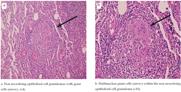
Figure 3. Lung biopsy. Histological examination
discussion
Sarcoidosis is a multisystem granulomatous disease; relatively rare in the pediatric popu-lation. Most of the reported cases of childhood sarcoidosis are accompanied by nonspecific cons-titutional symptoms as well as symptoms from particular organs such as lungs, eyes, skin, lym-ph nodes, liver and spleen. The lung is the most commonly involved organ in sarcoidosis. Bilateral hilar lymphadenopathy with or without lung involvement (state I and II) is the most common radiographic finding.1,2,3
The differential diagnosis of sarcoidosis depends largely on the clinical presentation of the disease. Granulomatous pulmonary infections especially those caused by mycobacteria and fungi should be ruled out. Neoplastic diseases, such as lymphoma, should be considered in cases with hilar adenopathy. Systemic-onset JRA should be in the differential diagnosis of early onset sarcoidosis.1
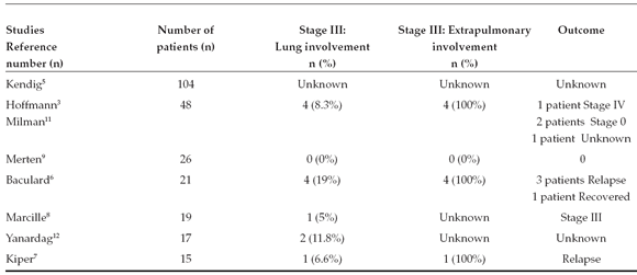
Kendig et al.1 published the largest patient group of childhood sarcoidosis with 104 patients. One hundred and three patients had BHL with or without lung involvement (stage I and II lung involvement).5 One patient in that study did not have hilar lymphadenopathy; whether this patient had any pulmonary involvement was not reported. Hoffman et al. reported 48 patients with childhood sarkoidosis from Denmark. In this report, although 8.3% had stage III lung involvement; all of them had extrapulmonary organ involvements as well.3 In the other case series of sarcoidosis there was no patient reported as state III lung involvement without any other organ involvement like ours 6,7,8,9 (Table 1).
Table 1. Childhood sarcoidosis cases
Eye, skin, musculosketelal features of sarcoidosis has been reported in 15% to 60% of children with sarcoidosis.1 None of these findings were present in our patient.
There are no specific blood tests for diagnosis of sarcoidosis. Laboratory evaluation may reveal elevated erythrocyte sedimentation rate or other acute phase reactants. Anemia, leukopenia, and eosinophilia are commonly seen. Immunological abnormalities include hypergammaglobulinemia and impaired delayed hypersensitivity on skin test. ACE is elevated in over 50% of children with late-onset sarcoidosis. Our patient’s laboratory test results were all within normal levels. BAL typically demonstrates an increased number of lymphocytes, most of which are activated helper-inducer T lymphocytes, that can cause a high CD4/CD8 ratio.1,2,3 Our patient’s BAL did not have increased lymphocytes, therefore, we could not evaluate CD4/CD8 ratio.
The diagnosis of sarcoidosis is confirmed by demonstrating a typical non-necrotising epithelioid cell granuloma on the biopsy specimen.1,2,3 Histological assessment of our patient's lung biopsy showed non-necrotising epithelioid cell granulomas, confirming the diagnosis of sarcoidosis.
Corticosteroids (CS) are the cornerstone of therapy for severe or progressive sarcoidosis (pulmonary or extrapulmonary), and often produce dramatic resolution of the disease. Other immunosuppressive agents, especially low-dose methotrexate (MTX) has been used to treat adult patients with sarcoidosis as a steroid-sparing agent.10 We treated our patient with CS and MTX and she became asymptomatic under this therapy. Prednisolone dose was reduced gradually to 2 mg/day in a year.
We presented an unusual patient showing that sarcoidosis can present only with lung involvement without hilar lympadenopathy and any other organ involvement. Sarcoidosis should be considered in the differential diagnosis of children with interstitial lung disease."
1. Shetty AK, Gedalia A. Childhood sarcoidosis: A rare but fascinating disorder. Pediatr Rheumatol on line 2008;23;6:16. [ Links ]
2. Mitchell DN, Scadding JG. Sarcoidosis. AM Rev Respir Dis 1974;110 (6):774-802. [ Links ]
3. Hoffman AL, Milman N, Byg KE. Childhood sarcoidosis in Denmark 1979-1994: incidence, clinical features and laboratory results at presentation in 48 children. Acta Paediatr 2004;93:30-6. [ Links ]
4. Cemlyn-Jones J, Gamboa F, Teixeira L, Bernardo J, et al. Sarcoidosis: a less common presentation. Rev Port Pneumol 2009;15(3):543-52. [ Links ]
5. Kendig EL, Niitu Y. Sarcoidosis in Japanese and American children. Chest 1980;77;514-6.
6. Baculard A, Blanc N, Boule M, Fauroux B, et al. Pulmonary sarcoidosis in children: a follow-up study Eur Respir 2001;17:628-35.
7. Kiper N, Anadol D, Ozcelik U, Gocmen A. Inhaled corticosteroids for maintenance treatment in childhood pulmonary sarcoidosis. Acta Pediatr 2001;90: 953-956. [ Links ]
8. Marcille R, McCarthy M, Barton JW, Merten DF, et al. Long-term outcome of pediatric sarcoidosis with emphasis on pulmonary status. Chest 1992 Nov;102(5):1444-9.
9. Merten DF, Kirks DR, Grossman H. Pulmonary sarcoidosis in children. Am Roentgenol 1980 Oct;135(4):673-9. [ Links ]
10. White ES, Lynch JP 3rd. Current and emerging strategies for the management of sarcoidosis. Expert Opin Pharmacother 2007;8:1293-1311. [ Links ]
11. Milman N, Hoffmann AL. Childhood sarcoidosis: long-term follow-up. Eur Respir 2008;31:592-8. [ Links ]
12. Yanardag H, Pamuk ON, Uygun S, Demirci S, et al. Sarcoidosis: child vs adult. Indian Pediatr 2006; 73:143-5. [ Links ]

