Servicios Personalizados
Revista
Articulo
Indicadores
-
 Citado por SciELO
Links relacionados
-
 Similares en
SciELO
  uBio
uBio
Compartir
Revista argentina de microbiología
versión impresa ISSN 0325-7541versión On-line ISSN 1851-7617
Rev. argent. microbiol. v.41 n.4 Ciudad Autónoma de Buenos Aires oct./dic. 2009
INFORME BREVE
Complete genome amplification of Equine influenza virus subtype 2
G. H. Sguazza1*, N. A. Fuentealba1, 2, M. A. Tizzano1, 3, C. M. Galosi1, 3, M. R. Pecoraro1.
1Cátedra de Virología, Facultad de Ciencias Veterinarias, Universidad Nacional de La Plata;
2Consejo Nacional de Investigaciones Científicas y Técnicas (CONICET);
3Comisión de Investigaciones Científicas de la Provincia de Buenos Aires (CIC), Argentina.
*Correspondence. E-mail: sguazza@fcv.unlp.edu.ar
Cátedra de Virología, Facultad de Ciencias Veterinarias, Universidad Nacional de La Plata 60 & 118, La Plata, CP 1900, Buenos Aires, Argentina
Telephone and fax number: 54 221 4257980
ABSTRACT
This work reports a method for rapid amplification of the complete genome of equine influenza virus subtype 2 (H3N8). A ThermoScriptTM reverse transcriptase instead of the avian myeloblastosis virus reverse transcriptase or Moloney murine leukemia virus reverse transcriptase was used. This enzyme has demonstrated higher thermal stability and is described as suitable to make long cDNA with a complex secondary structure. The product obtained by this method can be cloned, used in later sequencing reactions or nested-PCR with the purpose of achieving a rapid diagnosis and characterization of the equine influenza virus type A. This detection assay might be a valuable tool for diagnosis and screening of field samples as well as for conducting molecular studies.
Key words: Equine influenza virus; Genome; Diagnosis; RT-PCR.
RESUMEN
Amplificación del genoma completo del subtipo 2 del virus de la influenza equina. En este trabajo comunicamos un método rápido que permite la amplificación del genoma completo del subtipo 2 (H3N8) del virus de la influenza equina. Se utilizó la enzima transcriptasa reversa ThermoScriptTM en lugar de la transcriptasa reversa del virus de la mieloblastosis aviar o la transcriptasa reversa del virus de la leucemia murina de Moloney. Esta enzima ha demostrado tener una alta estabilidad térmica y la capacidad de hacer largas copias de ADN con una estructura secundaria compleja. El producto obtenido por esta técnica puede ser clonado y utilizado posteriormente en reacciones de secuenciación o de PCR anidada con la finalidad de lograr un diagnóstico rápido y la caracterización del virus de la influenza equina tipo A. Este ensayo de detección puede llegar a ser una valiosa herramienta para el diagnóstico y el análisis de muestras de campo, así como para la realización de estudios moleculares.
Palabras clave: Virus de la influenza equina; Genoma; Diagnóstico; RT-PCR.
Influenza viruses belong to the Orthomyxoviridae family and are classified into three great types: A, B, and C, according to antigenic differences in their nucleoprotein (NP) and matrix (M) proteins. Influenza viruses types B and C are predominantly human pathogens that have also been isolated from seals and pigs, respectively. On the other hand influenza viruses type A have been isolated from many species including humans, pigs, horses, marine mammals and a wide range of domestic and wild birds (10). Influenza viruses type A can be further subdivided in different serologically differentiated subtypes according to the structure and composition of two antigenic glycoproteins located in the surface of the virion: haemagglutinin (HA) involved in binding of the virus to host cells and neuraminidase (NA) implicated in release of virus from infected cells. At present, 16 HA and 9 NA subtypes have been identified among influenza A viruses. All these subtypes are found in avian species. However, only two equine influenza virus subtypes have been associated with the disease in horses, the H7N7 subtype (equi-1) and the H3N8 subtype (equi-2). The H7N7 subtype was first isolated from horses in Czechoslovakia in 1956 (prototype strain: A/equine1/Prague/56); later it was isolated in many countries of Europe and America. Although this subtype has not been isolated since 1980, it may still circulate in subclinical form and persist at very low levels in some parts of the world (10). The H3N8 subtype of equine influenza virus was first isolated in 1963 during an outbreak of this disease in Miami (prototype strain: A/equine2/ Miami/63) and can be found in different parts of the world, except in Australia, New Zealand, and Iceland. Recent studies of the H3N8 subtype of equine influenza viruses have demonstrated that these strains have diverged into two distinct evolutionary lineages (2).
The genome of equine influenza A virus contains eight linear segments of negative stranded RNA (about 13.6 kb). Six segments code for single proteins: the three viral polymerases (PA, PB1 and PB2), HA, NA and NP. The other two segments codify two proteins, one segment for matrix 1 and 2 (M1 and M2) and the other segment for non-structural protein 1 (NS1) and nuclear export protein (NEP) (5).
The analysis of virion RNA from influenza A strains indicates that all eight RNA segments contain a common sequence of 13 nucleotides at the 3' terminus and another common sequence of 12 nucleotides at the 5' terminus (3, 9). No homologies can be found among segments. On these bases, the aim of this work was to develop a rapid method that allows the genetic characterization of the whole genome of influenza virus using a procedure of reverse transcription followed by the amplification of all viral segments through polymerase chain reaction (RT-PCR).
The standard method for diagnosis of influenza is the isolation of influenza virus particles using embryonated chicken eggs or Madin-Darby canine kidney cells (MDCK). This system has the disadvantage of requiring 4-6 days for completion. The complete genome amplification by RT-PCR reported in this paper followed by subsequent amplicon sequencing is a more versatile method because it allows obtaining more detailed information about circulating strains and it will help in the virus surveillance.
To amplify the influenza virus genome, two primers based on the highly conserved non-coding sequences at the 5´ and 3´ end present in all viral genomic segments of influenza A virus were used, according to the consensus sequence regions previously reported (3, 9) and widely used to amplify the complete genome of human and avian influenza A virus (4) and equine influenza A virus subtype 1 (1). To confirm the presence of those conserved regions in the genome of equine influenza subtype 2, 50 different sequences of all segments from the viral genome retrieved from the GenBank at the National Center of Biotechnology Information (http://www.ncbi.nlm.nih.gov) were compared by multiple alignment using the CLUSTAL X (v1.8) software.
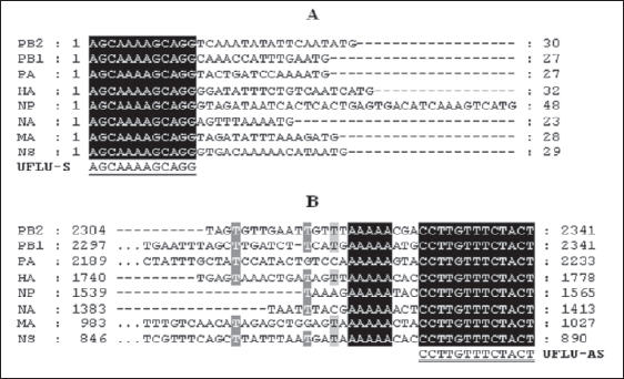
The consensus regions were identified as suitable sequences for use as primers in RT-PCR, and the oligonucleotide primers were subsequently commercially synthesized (Integrated DNA Technologies, Coralville, IA, USA). The primers used in this work were a 12-mer primer UFLU-S 5´AGCAAAAGCAGG 3´ and a 13-mer primer UFLU-AS 5´AGTAGAAACAAGG 3´ (Fig. 1).

Figure 1. Alignment of non-coding terminal regions of the eight segments of Influenza A virus (A/equine-2/Kentucky/5/02. GenBank accession numbers AY855340, AY855339, AY855338, AY855341, AY855342, AY855343, AY855344 and AY855345 respectively). The 5´ terminus of each segment has 12 conserved nucleotides (A), and the 3´ terminus has 13 conserved nucleotides (B).
An equine influenza virus strain, A/Equine-2/La Plata/ 2001 (H3N8), previously isolated in our laboratory during the year 2001, was used. The virus was propagated in the allantoic cavity of 11-day-old embryonated hen's eggs derived from a healthy flock.
Total RNA was extracted using Trizol reagent (GIBCO BRL, Gaithersburg, MD, USA). In brief, 500 μl of allantoic fluid (haemagglutination titre 1:128) were mixed with 500 μl of Trizol reagent. The mixture was extracted with 220 μl of chloroform. After centrifugation at 10000 x g for 10 minutes, the RNA in the aqueous solution was precipitated by adding an equal volume of isopropanol. The precipitated RNA was collected by centrifugation at 10000 x g for 20 minutes, washed by 70% ethanol, dissolved in 50 μl of RNAse-free water and measured by spectrophotometer (SmartSpec 3000, BIO-RAD, Hercules, CA, USA). RNA extracted from uninfected allantoic fluid was used as negative control. Total RNA and control RNA (375.06 ng/μl and 225.52 ng/μl respectively) were used for following steps.
One microgram of each RNA was used to produce a single-strand copy of DNA (ssDNA). This reaction was carried out using the avian myeloblastosis virus (AMV) reverse transcriptase (Promega Corporation, Madison, WI, USA), the Moloney murine leukemia virus (M-MMLV) reverse transcriptase (Promega) and the ThermoScript RT-PCR System kit (Invitrogen-Life Technologies, Buenos Aires, Argentina) under conditions specified by the suppliers, using a concentration 0.5 μM of the universal primer complementary to the conserved 3' end of all virus segments (UFLU-S).
The single-strand DNA (ssDNA) obtained with AMV, M-MLV and RT-ThermoScript was used directly as template in the PCR reaction whereas the cDNA generated by RT-ThermoScript was additionally employed for making a double-strand DNA copy (dsDNA) in agreement with the supplier's suggestion.
The second chain of DNA was synthesized using 3 μl of ssDNA previously obtained (approximately 100 ng) in a reaction mixture containing: 75 mM Tris-HCl (pH 8.8), 20 mM (NH4)2SO4, 0.01% Tween 20, 200 μM of each dNTPs, 1.5 mM of MgCl2 and 0.5 μM of UFLU-AS primer in a final volume of 24.5 μl. The mixture was incubated at 95 °C during 4 minutes, followed by the addition of 0.5 μl (2.5 U) of Taq DNA polymerase (Fermentas Inc, Glen Burnie, MD, USA) and it was incubated at 65 °C during 20 minutes.
The PCR reaction was performed by adding 5 μl from the ssDNA or dsDNA to a reaction mixture containing a final concentration of 75 mM Tris-HCl (pH 8.8), 20 mM (NH4)2SO4, 0.01% Tween 20, 200 μM of each one of dNTPs, 0.5 μM of each one of primers (UFLU-S and UFLU-AS), a variable concentration of MgCl2 (1, 1.5, 2 and 2.5 mM) and variable percentages of dimethil sulfoxide (0, 5 and 10% of DMSO) in a final volume of 49.5 μl. The mixture was heated at 95°C during 4 minutes and incubated with variable annealing temperatures (38, 40, 42 and 44 °C) for 2 minutes, then 0.5 μl (2.5 U) from Taq DNA polymerase (Fermentas) were added. The amplification program started with 1 cycle at 65 °C for 10 minutes, followed by 40 cycles at 92 °C for 2 minutes, the same annealing temperature initially used for 2 minutes, and 65 °C for 10 minutes; the program ended with one cycle at 72 °C for 10 minutes. The PCR product was run on agarose gel (1.5%) at 50 V during 6 hours, and the gel was stained with ethidium bromide and visualized with a UV transilluminator.
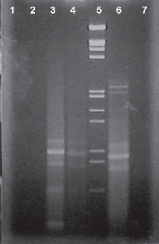
To evaluate the efficiency among reverse transcriptases AMV, M-MLV and ThermoScript, a protocol was used to amplify the whole genome of equine influenza virus. Improved results were observed when using dsDNA generated by ThermoScript-RT. When the ssDNA template obtained from AMV, M-MLV and ThermoScript was used, only a few bands could be observed in agarose gel (Fig. 2) corresponding to small size fragments of the genome.

Figure 2. RT-PCR using ssDNA as template: Lanes 3, 4 and 6: amplicon obtained from AMV, M-MLV and RT-ThermoScript; Lanes 1, 2 and 7: negative controls (RNA from uninfected allantoic fluid) obtained from AMV, M-MLV and ThermoScript RT, respectively. Lane 5: molecular size marker (λ EcoRI-HindIII Marker - Promega).
The optimum PCR reaction was obtained by adding 5 μl from the dsDNA to a reaction mixture containing a final concentration of 75 mM Tris-HCl (pH 8.8), 20 mM (NH4)2SO4, 0.01% Tween 20, 200 μM of each dNTP, 1.5 μM of MgCl2, 0.5 μM of each primer, 10% of DMSO and 2.5 U of Taq DNA polymerase.
An improved PCR amplification was carried out by heating the PCR mixture at 95 °C during 4 minutes, annealing at 40 °C for 2 minutes and then adding 0.5 μl (2.5 U) from Taq DNA polymerase (Fermentas). The amplification program started with 1 cycle at 65 °C for 10 minutes, followed by 40 cycles at 92 °C for 2 minutes, 40 °C for 2 minutes, and 65 °C for 10 minutes and a final extension at 72 °C for 10 minutes
The eight segments of equine influenza subtype 2 were amplified simultaneously using primers UFLU-S and UFLU-AS (Fig. 3). No amplified DNA was observed from the RNA negative control. Bands corresponding to basic polymerases (PB2 and PB1, segments 1 and 2 respectively) could not be clearly differentiated from each other because they were the same size (2341 bp each). It was followed by a band belonging to the acid polymerase (PA) of 2233 bp, segment 4 (HA) with 1778 bp, segment 5 (NP) of 1565 bp, segment 6 (NA) with 1413 bp, segment 7 (M) of 1027 bp and, at the end, a band belonging to segment 8 (NS) which showed an increased PCR product amount probably due to its smaller size.

Figure 3. RT-PCR using dsDNA as template: Lane 1: molecular size marker (λ EcoRI-HindIII Marker - Promega); Lane 2: RTPCR product.
In this study, the amplification conditions for the complete EIV genome were improved. The data presented in this paper demonstrate that this method could be a helpful tool in the surveillance of the equine influenza virus and appropriate for cloning the eight genomic segments, which will facilitate large-scale EIV genome sequencing and greatly ease systematic genetic analyses of the virus. Adeyefa et al. (1) have described a multiplex RTPCR method in which 12-mer and 13-mer oligonucleotides complementary to the conserved regions were used, resulting in simultaneous amplification of all of the eight RNA segments of Equine Influenza virus subtype 1. In this work, a ThermoScript RT instead of AMV or M-MLV RT was used to amplify the full length genome of equine influenza virus subtype 2 (H3N8). This enzyme has higher thermal stability and is described as suitable to make long cDNA with complex secondary structure.
The product obtained by this method can be cloned and used in later sequencing reactions or nested-PCR with the purpose of achieving a rapid diagnosis and characterization of the equine influenza virus type A (1, 8).
This test is much faster than the isolation of the virus from nasopharyngeal swabs/washings using embryonated eggs or cell cultures and the possibility of automation will allow handling a large number of clinical samples more conveniently than with serological tests (7, 8).
RT-PCR might be useful in detecting inapparent, subclinical infection in animals that contribute to the spread of the virus. Accurate identification of infected animals and inapparent carriers is essential for the control of this disease.
1. Adeyefa CA, Quayle K, McCauley JW. A rapid method for the analysis of influenza virus genes: application to the reassortment of equine influenza virus genes. Virus Res 1994; 32: 391-9. [ Links ]
2. Daly JM, Lai AC, Binns MM, Chambers TM, Barrandeguy M, Mumford JA. Antigenic and genetic evolution of equine H3N8 influenza A viruses. J Gen Virol 1996; 77: 661-71. [ Links ]
3. Desselberger U, Racaniello VR, Zazra JJ, Palese P. The 3' and 5'-terminal sequences of influenza A, B and C virus RNA segments are highly conserved and show partial inverted complementarity. Gene 1980; 8: 315-28. [ Links ]
4. Hoffmann E, Stech J, Guan Y, Webster RG, Perez DR. Universal primer set for the full-length amplification of all influenza A viruses. Arch Virol 2001; 146: 2275-89. [ Links ]
5. McCauley JW, Mahy BW. Structure and function of the influenza virus genome. Biochem J 1983; 211: 281-94. [ Links ]
6. Oxburgh L, Klingeborn B. Cocirculation of two distinct lineages of equine influenza virus subtype H3N8. J Clin Microbiol 1999; 37: 3005-9. [ Links ]
7. Oxburgh L, Hagström A. A PCR based method for the identification of equine influenza virus from clinical samples. Vet Microbiol 1999; 67: 161-74. [ Links ]
8. Quinlivan M, Cullinane A, Nelly M, Van Maanen K, Heldens J, Arkins S. Comparison of sensitivities of virus isolation, antigen detection, and nucleic acid amplification for detection of equine influenza virus. J Clin Microbiol 2004; 42: 759-63. [ Links ]
9. Skehel JJ, Hay AJ. Nucleotide sequences at the 5' termini of influenza virus RNAs and their transcripts. Nucleic Acids Res 1978; 5: 1207-19. [ Links ]
10. Webster RG, Bean WJ, Gorman OT, Chambers TM, Kawaoka Y. Evolution and ecology of influenza A viruses. Microbiol Rev 1992; 56: 152-79. [ Links ]
Recibido: 28/04/09
Aceptado: 22/09/09


