


Services on Demand
Journal

Article
 English (pdf)
 Article in xml format
 Article references
 Send this article by e-mail
Indicators
-
 Cited by SciELO
Related links
-
 Similars in
SciELO
Similars in
SciELO  uBio
uBio
Share

 Permalink
Revista argentina de microbiología
Print version ISSN 0325-7541On-line version ISSN 1851-7617
Rev. argent. microbiol. vol.48 no.2 Ciudad Autónoma de Buenos Aires June 2016
http://dx.doi.org/10.1016/j.ram.2016.03.006
BRIEF REPORTS
http://dx.doi.org/10.1016/j.ram.2016.03.006
LED fluorescence microscopy in the diagnosis of tuberculosis: Fading and restaining of smears for external quality assessment
Microscopía de fluorescencia con lámpara LED en el diagnóstico de tuberculosis: decoloración y recoloración de extendidos para evaluación externa de calidad
Sonia Allassiaa, Mónica Aranibarb, Mónica Boutonneta, Viviana Caseríoa, Ana Alicia Etchartb, Sandra Fajardoa, Mónica Garcíac, Noemí Gomezd, Alba Marisa Guniad, María Virginia Gustincice, Viviana Izquierdob, Arnaldo Andrés Jarad, Graciela Kozickya, Mario Matteoc, Carlos Pellegrinia, Silvia Pellegrinod, Sebastián Pérez Cataláne, Susana Poggic, Carina Sacramoned, Gabriela María Santisoe, Alejandro Soutoc, Ana María Tognerie, Lidia Wolfff, Sandra Vilcheg, Daniel Elettig, María Susana Imazg, *
a. Dirección de Bioquímica de la Municipalidad de Rosario, Rosario, Santa Fe, Argentina
b. Laboratorio de Tuberculosis, Hospital San Roque, San Salvador de Jujuy, Jujuy, Argentina
c. Laboratorio de Tuberculosis, Hospital F.J. Muñiz, CABA, Argentina
d. Laboratorio de Tuberculosis, Laboratorio Central de Salud Pública, Resistencia, Chaco, Argentina
e. Laboratorio de Bacteriología, Hospital Interzonal General de Agudos «Evita», Lanús, Buenos Aires, Argentina
f. Laboratorio de Bacteriología, Hospital Rawson, Córdoba, Argentina
g. Departamento Diagnóstico y Referencia, Instituto Nacional de Enfermedades Respiratorias «Emilio Coni», ANLIS «C.G. Malbrán», Santa Fe, Argentina
Received 18 December 2015; accepted 22 March 2016
Available online 10 June 2016
* Corresponding author. E-mail address: suimaz@yahoo.com (M.S. Imaz).
0325-7541/© 2016 Asociacón Argentina de Microbiología. Published by Elsevier España, S.L.U. This is an open access article under the CC BY-NC-ND license (http://creativecommons.org/licenses/by-nc-nd/4.0/).
Abstract
Blinded rechecking is a method proposed for external quality assurance (EQA) of auramine-stained acid-fast bacilli (AFB) smears using fluorescence microscopy (FM), however, this procedure is not well developed and slides fading over time could compromise its implementation. Since bleaching of fluorescent molecules involves temperature-dependent chemical reactions, it is likely that low temperatures could slow down this process. We stored auramine-stained slides under different environmental conditions, including −20 °C, and examined them over time. The slides stored in all the environments faded. At −20 °C, fading was not reduced in relation to room temperature. Restaining and re-examining smears after five months showed that the slides containing saliva and storage at −20 °C were associated with failure in AFB reappearance. In conclusion, the practice of freezing slides until they are viewed should be discouraged as it has a negative effect on blinded rechecking by reducing reading concordance after restaining. Specimen quality should be considered when interpreting FM-EQA results.
Keywords
Tuberculosis; LED fluorescence microscopy; External quality assessment.
Resumen
La relectura usando microscopía fluorescente con lámpara LED es una metodología propuesta para la evaluación externa de calidad (EEC) de los extendidos teñidos con auramina empleados para detectar bacilos ácido-alcohol resistentes (BAAR), pero el procedimiento está parcialmente desarrollado y la decoloración de los BAAR con el transcurso del tiempo puede comprometer su implementación. La decoloración de moléculas fluorescentes involucra reacciones químicas temperatura-dependientes, por lo que la reducción de la temperatura podría enlentecerla. Guardamos extendidos coloreados en distintas condiciones, incluyendo a −20 °C, y los examinamos a distintos tiempos. Las láminas guardadas en todos los ambientes se decoloraron; a −20 °C la decoloración fue más rápida que a temperatura ambiente. La recoloración evidenció que en extendidos de saliva o conservados a −20 °C existía mayor probabilidad de que los BAAR no reaparecieran. En conclusión, la conservación de extendidos en freezer debe evitarse, ya que reduciría la concordancia de lectura luego de recolorear para la EEC. La calidad de la muestra debe considerarse para interpretar la EEC.
Palabras clave
Tuberculosis; Microscopía de fluorescencia LED; Evaluación externa de calidad.
In comparison with Ziehl Neelsen (ZN) microscopy, conventional (mercury vapor lamp) fluorescence microscopy (FM) using auramine O staining, can detect approximately 5-10 % more acid-fast bacilli (AFB) positive smears7; however, its use has been limited due to the requirement of expensive mercury vapor lamps and dark room facilities. The advent of light-emitting diode (LED) technology, which is inexpensive and uses a long lifespan lamp, has led the World Health Organization (2011) to recommend the use of LED-FM as an alternative to ZN in a phased manner10. However, appropriate rechecking procedures and External Quality Assessment (EQA) policies for FM are not well developed. The fading of stains is generally believed to occur much faster with auramine O than with ZN stained slides, particularly after light exposure5. This phenomenon is attributed to the photo-oxidation property of auramine O as a fluorescent dye. Therefore, the restaining of slides seems to be mandatory to avoid gross errors by controllers during the rechecking process of FM, the most effective method of proficiency testing of sputum smears examined for acid-fast bacilli (AFB) in peripheral centers.
Photobleaching mechanisms of organic dyes, such as auramine O, are complex and mostly unknown. All fluorescent organic molecules photobleach sooner or later at room temperature (RT). Since photobleaching involves temperature-dependent chemical reactions, it is likely that low temperatures could considerably slow down the process, and, possibly, because of this observation, freezing slides until they are viewed by FM is a common practice. Nevertheless, to our knowledge, the auramine O fading effect under this low temperature has not yet been proved. Furthermore, even when bleaching of auramine O-stained smears has been studied in regard to the speed of fading under different environmental conditions of storage3, no study has yet determined whether these maintenance conditions may affect the reading of the stored slides after restaining. In this work we evaluated the effect of different storage temperatures, including −20 °C, on the fading of auramine O-stained smears. Furthermore, the storage conditions and other possible interfering factors were taken into account in order to evaluate the reading concordance of the stored smears after restaining.
The present study was conducted as part of a multicenter study developed to assess the feasibility of using LED-FM in peripheral laboratories in Argentina. For the purpose of this multicenter study, FM slides were examined under Olympus CX31 microscopes with TK-LED illumination (Tolket, Argentina), using 200× magnification for screening and 400× magnification for confirming and quantifying bacillary counts in the slides. Slides were prepared for staining in duplicate using the hot ZN technique (0.3 % carbol fuchsin and 0.1 % methylene blue) or with 0.1 % auramine O, counterstained with 0.5 % potassium permanganate, for LED-FM. Two lengths of 200× magnification were read before the slides were declared negative. A subset of 296 diagnostic sputum specimens slides, identically scored by two microscopists on initial reading, was selected for storage under a variety of conditions. Smears differed in specimen quality and bacillary content. The slides were allocated in closed slide boxes to one of four storage environments, in darkness, as follows: (i) in freezer (−20 °C), (ii) in refrigerator (4 °C), (iii) at RT (22 °C) and (iv) in an incubator at 30 °C. The subsets of 74 slides stored in each of the 4 environments were composed of 10 negative slides and 64 positive smears that were selected in order to enrich the positive panel with 50 % of low positive smears (scanty and positive (+)), as follows: 16 scanty, 16 positive (+), 20 positive (++) and 12 positive (+++) slides. This high proportion of low positive smears was included in an effort to target the slides that were most at risk of being missed during the incubation period due to the fading effect at different environments. AFB were counted at the start of the trial and at monthly intervals in a blinded manner. When no AFB could be identified any longer by the microscopist, the slide was subjected to a second controller, who was not blinded to the original result, in order to confirm the maximum of positivity; furthermore, when no bacillus was found in the first two lengths of 200× magnification, two additional lengths were read by this second controller until the slides were declared negative. Even if AFB were not detected by this second controller, the slide continued to be examined during the following month in order to confirm its fading. Only those smears that stayed negative after two consecutive months were considered to have faded.
Table 1 shows the fading rate of fluorescent staining slides with different grades of positivity, considering all smears kept under all temperature conditions. As previously reported3, the fading speed significantly differed between the slides containing "numerous AFB" (positive ++ and +++) and those containing "less numerous AFB" (scanty and positive +) as could be observed by the analysis of the linear regression slope; the proportion of positive smears that faded to negative was 10.40 %/month (CI - 8.81; 12.02) in weakly positive slides (scanty and positive +) whereas it dropped only to 2.60 %/month (CI - 1.61; 3.62) in smears with a higher quantity of AFB (positive ++ and +++). By month 3, almost 30 % of the "less numerous AFB" slides were negative, whereas this proportion reached less than 5 % in those slides with numerous AFB. Another subsets of slides (composed of 24 slides with 4 negative smears and a similar proportion of smears having different grades of positivity as previously described) were kept under the four previously described environments until the third month, when smears were examined for the first time; of 40 strong positive slides, 38 (95 %) remained positive whereas this proportion reached 72.50 %% (29/40) in those slides with "less numerous AFB"; these proportions did not significantly differ from those obtained with slides read at monthly intervals (96.10 % and 72.66 % for "numerous AFB" and "less numerous AFB" slides, respectively, p < 0.05) (Table 1). Moreover, we found that weakly positive slides began to fade during the first month of observation; however, according to Xia et al.11 and Radhakrishnan et al.6, the proportion of fading of fluorescence-stained smears reported by us with weakly positive smears (10 %/month) was considerably lower than that described by Minion et al.3, who found a fading speed of about 25 %/month. This difference could be attributed to the limitations of the device used by Minion et al.2, who informed that the Lumin attachment was difficult to focus and showed lower excitation light intensity than other devices, which might limit the identification of those AFB that were slowly losing their intensity over time.
Table 1. Influence of storage time on the fading rate of auramine O-stained slides
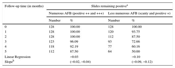
a - Slides undergoing storage in different environments were combined.
b - Intercept forced through 1.0. The values in parentheses represent 95 % confidence intervals.
Contrary to our thoughts, when considering all the slides with "less numerous AFB" that originally read as positive (combined scanty and positive +), the fading at the lowest temperature of −20 °C was even more rapid than at RT. AFB kept at RT seemed to disappear significantly more slowly than in all the other conditions, as could be observed by analyzing the confidence intervals of the slopes obtained after linearizing the fading rate of fluorescent stained slides (Table 2). Two circumstances could accelerate photobleaching: the first one is working at room temperature, as fading increases with temperature because more reactions involved in the process become activated. The second one is the water or water-like environment, because in aqueous solutions, fluorophores are easily attacked by small reactive molecules such as oxygen or water itself13. This last phenomenon could explain why, contrary to our expectations, at a low temperature of −20 °C, fading was not reduced in relation to RT; water condensation accumulated on the cold smears after removal from the freezer could accelerate fading more than constant ambient humidity did. Nevertheless, the explanation is not clear as this phenomenon could not explain the fact that the fading effect could be observed even immediately after the first defrost done after one or three months of storage.
Table 2. Fading of low positive slides (scanty and positive +) under different storage conditions

a - Intercept forced through 1.0. The values in parentheses represent 95 % confidence intervals.
As fading occurred with stored auramine O-stained smears, restaining all the slides would be the only way to maintain the efficiency of blinded rechecking. Therefore, in order to mimic the procedure that would be followed for blinded rechecking before the first control, we decided to restain all the stored smears after five months of storage. FM readings were done by the same microscopists in a blinded manner. False negative (FN) results (positive at the initial reading and negative after restaining) were subjected to a second control reading, following the previously described reading scheme, aiming for the maximum confirmation of positive results. A result was defined as true-positive if it was confirmed at rechecking and a false-negative was declared only when the slide was positive in the original reading but no AFB could be found after restaining. After the rereading process, the slides were stained with 0.1 % methylene blue for 1 min in order to assess the sputum quality microscopically. Saliva was defined as any specimen containing a predominance of squamous epithelial cells, without the presence of mucus.
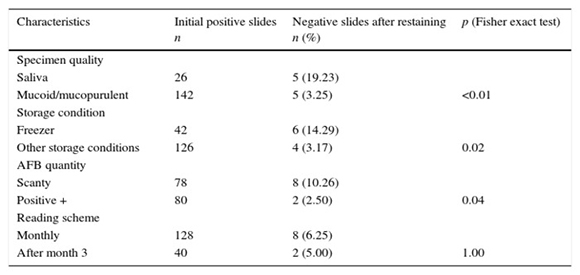
The concordance analysis after restaining showed that 326 of the 336 initially positive slides displayed results consistent with the original reading. All the 10 slides that resulted negative had low grade of positivity (scanty or positive +). The analysis of the factors associated with the loss of visible AFB in this group of 168 low positive slides, showed a significantly increased proportion of discordant results in the saliva specimens, scanty smears and frozen storage slides (Table 3). No statistically significant difference in the proportion of positive slides after restaining was observed between the slides read at monthly intervals and those examined at month 3. After controlling by confounders by multivariate logistic regression, only the smears made from saliva specimens and storage at −20 °C were negatively associated with the concordant results after restaining, with OR being 0.17 (0.04, 0.72), and 0.17 (0.04, 0.73) respectively. Similarly, Van Deun and Portaels9 showed a loss of AFB in ZN-restained smears made from liquefied sputum but no such losses could be shown in smears made from fresh sputum; they hypothesized that under this latter condition, AFB removal during the restained process might be prevented as the bacilli were presumably safe in a matrix of mucoid material. In the same way, water condensation accumulated on the cold smears after removal from the freezer could affect AFB fixation, predisposing the bacilli to be washed out of the smears during restaining. Negative results may also occur during cross checking of low grade of positivity since rare AFB, even without fading, do not always reappear due to the inherently less reproducibility involved in the examination of low positive slides. Nevertheless, in the present work, in order to reduce this variability during the rechecking process, special efforts were made to confirm low positivity slides by including a non-blinded second controller who, in cases of apparent FN, extended the number of fields examined. Furthermore, even if this lack of reproducibility among low positive score slides could add variability to the reading, this should not affect the differences of concordance observed among the slides kept at various environments or made from specimens of different quality.
Table 3. Univariate analysis of specimen quality, bacillary load, storage conditions and reading schemes associated with loss of positivity after restaining 168 low positive slides
Furthermore, even when we take into account those low positive smears kept out of freezer storage conditions, about 16 % of saliva specimens, which apparently lost AFB during restaining (data not shown), gave false negative results. In practice, during the blinded rechecking process, these discordances would cause "false positive results" (a negative smear declared by the controller that is misread as positive by the technician at the periphery laboratory), as the controller, after restaining the slide, would not be able to observe those AFB that were observed by the laboratory workers at the periphery center. It is well known that AFB positivity in smear microscopy is closely related to the quality of the specimen1,4 Hence, in Argentina, the results of 20 years of experience in EQA by blinded rechecking showed that, in a large random sample, the proportion of positive slides made from saliva samples was only about 7 %1. Applying our 16 % rate of discordant results found in saliva specimens after restaining to the observed 7 % frequency of saliva among positive smears reported by Kusznierz et al.1, the restaining process previous to rechecking might thus add 1.1 % false-positives at most. Low false positive (LFP) errors are to be expected, since AFBs are not homogeneously distributed in sputum. For these reasons, the interpretation of LFP errors may be considered separately from major high false positive/high false negative errors8. In practice, occasional scanty false-positive results should simply be disregarded for analysis of cross checking results.
Contrary to what Yip et al. described12, our readers did not find that restained slides were harder to read. Although unclear, we hypothesized that this difference in restaining performance may be associated with a higher concentration of auramine O and a lower proportion of the potassium permanganate employed in the staining and counterstaining solutions, respectively in the study described by Yip et al. in comparison to ours.
In conclusion, restaining all the smears before rechecking, a procedure that has proved essential to reach valid evaluations for routine FM external quality assessment, could be performed easily as usual. The common practice in FM of refrigerating slides until they are viewed should be discouraged as this storage condition has negative effects on blinded rechecking both by accelerating the fading rate (in comparison with RT) and, also, in the case of freezer conditions, by reducing the reading concordance of stored slides after restaining. Furthermore, smears made from saliva specimens were significantly associated to "false false positive results" on cross-checking, which may happen when AFB that were weakly trapped in a non-mucoid medium are washed out of the smears during the restaining process. Nevertheless, considering that the proportion of positive slides of saliva in the Argentinean laboratory network seems to be low, the negative effect of saliva specimens on the efficiency of the rechecking process appears to be low, especially in the context of other limitations of the cross checking process previously described9. Saliva specimens must continue to be systematically processed, since, although positivity in these specimens is lower than that with good quality sputa, their examination allows the diagnosis of additional TB cases.
Our study confirms that restaining all the auramine O-stained smears is necessary before re-examination; however, this process requires much more additional work, a major expense for laboratories and may cause new problems that could affect the accurate assessment of LED-FM. Thus, blinded rechecking programs continue to be a challenge for FM, encouraging the importance of further research to evaluate alternative methodologies for external quality assessment.
Ethical disclosures
Protection of human and animal subjects
The authors declare that no experiments were performed on humans or animals for this investigation.
Confidentiality of data
The authors declare that no patient data appears in this article.
Right to privacy and informed consent
The authors declare that no patient data appears in this article.
Funding
This study was subsidy supported by Agencia Nacional de Promoción Científica y Tecnológica PAE-PID-2007-00127.
Conflict of interest
The authors declare that they have no conflicts of interest.
1. Kusznierz GF, Latini OA, Sequeira MD. Quality assessment of smear microscopy for acid-fast bacilli in the Argentine tuberculosis laboratory network, 1983-2001. Int J Tuberc Lung Dis. 2004;8:1234-41. [ Links ]
2. Minion J, Pai M, Ramsay A, Menzies D, Greenaway K. Comparison of LED and conventional fluorescence microscopy for detection of acid fast bacilli in a low-incidence setting. PLoS One. 2011;6:e22495. [ Links ]
3. Minion J, Shenai S, Vadwai V, Tipnis T, Greenaway C, Menzies D, Ramsay A, Rodrigues C, Pai M. Fading of auramine-stained mycobacterial smears and implications for external quality assurance. J Clin Microbiol. 2011;49:2024-6. [ Links ]
4. Pollak L, Urbanczik R. La relación entre la calidad de la muestra y la positividad en microscopía. Bol Inform Inst Nac Tuberc. 1969;2:5-8. [ Links ]
5. Poulios I, Avranas A, Rekliti E, Zouboulis A. Photocatalytic oxidation of Auramine O in the presence of semiconducting oxides. J Chem Technol Biotechnol. 2000;75:205-12. [ Links ]
6. Radhakrishnan R, Prabuseenivasan S, Balaji S, Sankar U, Thomas A, Kumar V, Selvakumar N. Blinded rechecking of acid-fast bacilli smears by light-emitting diode microscopy. Int J Tuberc Lung Dis. 2013;17:1220-3. [ Links ]
7. Steingart KR, Henry M, Ng V, Hopewell PC, Ramsay A, Cunningham J, Urbanczik R, Perkins M, Aziz MA, Pai M. Fluorescence versus conventional sputum smear microscopy for tuberculosis: a systematic review. Lancet Infect Dis. 2006;6:570-81. [ Links ]
8. Torrea G, Chakaya J, Mayabi M, Van Deun A. Evaluation of the Fluoreslen S and fluorescence microscopy blinded rechecking trial, Nairobi, Kenya. Int J Tuberc Lung Dis. 2008;12:658-63. [ Links ]
9. Van Deun A, Portaels F. Limitations and requirements for quality control of sputum smear microscopy for acid-fast bacilli. Int J Tuberc Lung Dis. 1998;2:756-65. [ Links ]
10. WHO. Fluorescent Light-Emitting Diode (LED) Microscopy for Diagnosis of Tuberculosis: Policy Statement. Geneva: World Health Organization; 2011. WHO/HTM/TB/2011.8. [ Links ]
11. Xia H, Song YY, Zhao B, Kam KM, O'Brien RJ, Zhang ZY, Sohn H, Wang W, Zhao YL. Multicenter evaluation of Ziehl-Neelsen and light-emitting diode fluorescence microscopy in China. Int J Tuberc Lung Dis. 2013;17:107-12. [ Links ]
12. Yip CW, Chan MY, Cheung WF, Yu KW, Tang HS, Kam KM. Random blinded rechecking of sputum acid-fast bacilli smear using fluorescence microscopy: 8 years' experience. Int J Tuberc Lung Dis. 2012;16:398-401. [ Links ]
13. Zondervan R, Kulzer F, Kolćhenko MA, Orrit M. Photobleaching of Rhodmine 6G in poly (vinyl alcohol) at the ensemble and single-molecule levels. J Phys Chem. 2004;108:1657-65.

