


 Inglés (pdf)
 Articulo en XML
 Referencias del artículo
 Enviar articulo por email
 Citado por SciELO
  Similares en
SciELO
Similares en
SciELO  uBio
uBio 
 Permalink
The behavior of Trichoderma afroharzianum strain Th2RI99 confronted to Rhizoctonia solani AG4-HG II in dual culture was observed under light (LM) and scanning electron micro-scope (SEM). There is evidence of mycoparasitism in the Trichoderma harzianum complex, but no report is available regarding T. afroharzianum, since it has been just recently described as a new species, previously identified with phy-logenetic analysis with 3 genes1.
Two mycelial plugs (5mm diam.), from Th2RI99 and the pathogens Bipolaris sorokiniana, Fusarium graminearum, Sclerotium minor and R. solani cultures (grown in potato dextrose agar for 72 h at 25 °C in darkness), were placed 3cm apart from each other on 1.2% water agar plates in independent assays. Three replicates were incubated and observed at 24, 46, 47, 48 and 72h. Rectangular sections from the confrontation area were mounted on slides in 3% KOH, stained with 10% lactophenol blue or 5mM white calcofluor (Fluka, Sigma-Aldrich) for 5min and washed with distilled water2,4,5. The observation of samples under LM (Olympus BX51) coupled to a digital camera (Cool Snap-Pro) was carried out using bright field, differential contrast inter-ference and epifluorescence (360-370 nm). Digital images were taken using Image-Pro Solution software (Media Cybernetics).
Samples for SEM observation were prepared following3. Only samples against R. solani showed signs of mycoparasitism. At46and 47h, multiple coilingsfrom Th2RI99 hyphae around R. solani mycelium were evident (Figs. 1A and B). At 48 h, the cell wall of R. solani presented the formation of a pore, caused by the penetration tube of Th2RI99 (Fig. 2A).
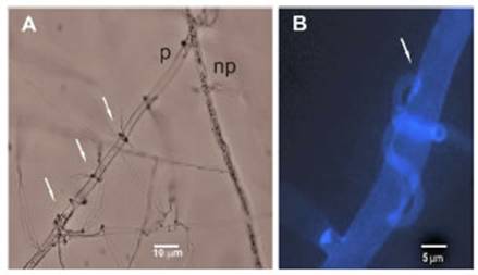
Figure 1: Light microscope observation of the interaction between hyphae of Trichoderma afroharzianum Th2RI99 (T) and Rhizoctonia solani AG4-HG II (R) in dual culture (WA 1.2%) after 47 h. (A) Arrows: coilings of Th2RI99 on R. solani; p: plasmolized hyphae; np: not plasmolized hyphae. (B) Calcofluor dye. Arrow: detail of coiling of Th2RI99 on R. solani.
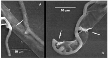
Figure 2: Scanning electron microscope observation of the interaction between hyphae of Trichoderma afroharzianum Th2RI99 (T) and Rhizoctonia solani AG4-HG II (R) in dual culture (WA 1.2%) after 48 h. (A) Arrow: formation of penetration pore in hypha of R. solani. (B) Arrows: Th2RI99 penetration tubes around the apical hypha of R. solani.
In one of the mycoparasitism events, the apical zone of a R. solani hypha had up to 5 penetration tubes from the antagonist (Fig. 2B).
This is the first report of mycoparasitic interaction between T. afroharzianum and R. solani.
Conflicts of interest
This work has been carried out as a Technological Association Agreement with Rizobacter S.A.
Acknowledgements
We thank Disciplinary Project 2019-PD-E4-I069-001 (INTA) and the Technological Association Agreement INTA-Rizobacter S.A. for funding this work.
Received 11 May 2022
accepted 27 September 2022

