


Services on Demand
Journal

Article
 English (pdf)
 Article in xml format
 Article references
 Send this article by e-mail
Indicators
-
 Cited by SciELO
Related links
-
 Similars in
SciELO
Similars in
SciELO  uBio
uBio
Share

 Permalink
Biocell
Print version ISSN 0327-9545
Biocell vol.29 no.3 Mendoza Aug./Dec. 2005
Construction and application of a yeast expression system for thymosin a1
Chen Feng*, Chen Xiang-Ming**, Chen Zhi*, Jiang Han-Liang*, Pan Xiao-Ping*, Hu Zhong-Rong*, Liu Rong-Hua* and Chen Xiao-Ming*
* Institute of Infectious Diseases, 1st Affiliated Hospital, Medical College of Zhejiang University, Key Lab. of Infectious Diseases, Ministry of Public Health, Hangzhou, 310003, CHINA.
** Department of Anesthesiology, 2nd Affiliated Hospital, Medical College of Zhejiang University, Hangzhou, 310009, CHINA.
Address correspondence to: Dr. Feng Chen. The Institute of Infectious Diseases, 1st Affiliated Hospital, Medical College of Zhejiang University, 79 Qingchun Road, Hangzhou, 310003. CHINA. Fax: (+86-571) 87068731. E-mail: xinlanchen222@hotmail.com
ABSTRACT: We want to construct a yeast expression system for thymosin a1 (Ta1) to make the orally administered Ta1 preparation possible. The whole Ta1 DNA fragment was obtained by PCR. After being digested with restriction enzymes, it was cloned into pYES2 vector. Sequencing was performed to identify the recombinant. The sequence of Ta1 in recombinant coincided with the original one reported in Genbank. When pYES2-Ta1 plasmid was transformed into yeast, galactose instead of glucose was used to induce Ta1 expression. Western blot was performed to identify the quality of the expressed Ta1. Dried yeast containing pYEST2-Ta1 was fed to Balb/c mice whose immunities were inhibited by cyclophosphamide in advance. Synthesized Ta1 peptide was used as positive control and empty yeast was used as negative control. Compared with the negative control group, both dried yeast containing pYEST2-Ta1 and synthesized Ta1 peptide can significantly increase the CD8+ level (22.74±1.09 and 18.77±4.72 vs 7.49±2.14, p<0.01), while both of them had little effect on the CD4+ lymphocytes (61.86±6.94 and 65.91±4.78 vs 57.93±10.40, p>0.05). We concluded that a high effective yeast expression system for Ta1 was constructed successfully and the Ta1 protein expressed by this system can improve CD8+ level in immune inhibited mice.
Keywords: Thymosin a1. Yeast expression system. Biological activity. Orally administered.
Introduction
Thymus is a vital immune organ and plays a very important role in the development and maintenance of lymph system. The studies on thymosins were initiated in 1965. Thymosins extracted from animal thymus had been used as an adjunct drug in patients with carcinoma, viral infection and immunodeficiency in clinical practice. However, the extract containing miscellaneous animal proteins may easily lead to allergy in patients. Further investigation implied that the extract from bovine thymus gland can be divided into five groups. Among them, thymosin fraction 5 has the most ability to improve immune response. It turned out that thymosin fraction 5 consisted of a mixture of small polypeptides, such as Ta1, polypeptide β1, prothymosin a, parathymosin, and thymosin β4 and so on. They are biologically important peptides with diverse intracellular and extracellular functions (Hannappel and Huff, 2003). Ta1 is the most potent ingredient in thymosin and the biological activity of pure Ta1 is 10~1000 times over that of total thymus extract.
Ta1, a 28-amino acid peptide, is an immune modifier that has been shown to trigger maturational events in lymphocytes, to augment T-cell function, and to promote reconstitution of immune defects (Ancell et al., 2001). Ta1 has been shown to promote disease remission and cessation of hepatitis B virus (HBV) replication in patients with HBeAg-positive chronic hepatitis B without significant side effects (Mutchnick et al., 1991; Chien et al., 1998; Saruc et al., 2003). The efficacy and safety of IFN-a 2a and Ta1 combination therapy in patients with chronic hepatitis C were determined. The combination therapy produced a good response rate and minor side effects, especially in naive patients with acceptable safety profile (Kullavanuaya et al., 2001). Moreover, clinical trials using Ta1 in the treatment of patients with immunodeficiency or cancer indicate that this agent is nontoxic, enhances immune responsiveness and augments specific lymphocyte functions, including lymphoproliferative responses to mitogens, maturation of T cells, antibody production, and T cell mediated cytotoxicity. (Moody et al., 2002; Rasi et al., 2003).
Pure chemically synthesized Ta1 has already been used as an immune-mediate factor in clinical practice for the treatment of HBV and hepatitis C virus (HCV) infections, non-small cell lung cancer, hepatocellular carcinoma, AIDS and malignant melanoma. Ta1 is able to potentiate the action of cytokines and also reduce the hematological toxicity of cytotoxic drug therapy (cyclophosphamide-, 5-fluorouracil-, dacarbazine- or ifosfamide-based regimens). It was in phase III trials in the US in combination with PEGylated IFN-a, and it has been launched in Argentina, China, Peru, the Philippines and Singapore for the treatment of chronic HBV infection (Billich, 2002). However, high price and administration by inconvenient intramuscular injection have limited its wide use. A new approach is needed to produce high qualitative, cheap and orally administered Ta1 to satisfy clinical practice. In this study, Ta1 was cloned into a yeast expression vector and then the yeast with its expressed peptides was fed to Balb/c mice, whose immunities were inhibited by cyclophosphamide in advance. The effects of Ta1 on the immune function of the mice were observed.
Material and methods
Construction of T a1 clone
Obtainment of the whole Ta1 gene: The whole Ta1 single DNA chain and primers were synthesized by Shanghai Boshide Corporation according to the Genbank sequence (E00106). Up stream primer is 5'GGA AGC TTT CTG ATG CTG CTG TTG 3', which contains Hind III restriction enzymatic site, and down stream primer is 5' TAT GGA TCC TAG TTC TCA GCC TCT TCG 3', which contains BamH I restriction enzymatic site. The whole Ta1 double DNA chains were obtained by PCR. Initial denaturation was at 95ºC for 2min followed by 30 cycles at 94ºC (1min), 54ºC (1min) and 72ºC (1min) in an Eppendorf PCR machine. The final elongation step was at 72ºC for 10min. High fidelity Pwo DNA polymerase (Cat. 1644947) was purchased from Roche Corporation.
Digestion: Hind III (Cat. R6041) and BamH I (Cat. R6021) enzymes were purchased from Promega Corporation. Ta1 PCR products were digested with both Hind III and BamH I restriction enzymes at 37ºC for 1h, so was the pYES2 plasmid (ClonTech Corporation, Cat. V825-20). The digested products were reclaimed from agarose gels, and then they were purified with the QIAquick Gel Extraction Kit (Qiagen Corporation, Cat. 28704).
Ligation: The enzymatic digestive products of Ta1 PCR product and pYES2 plasmid were ligated by T4 ligase (Roche Corporation, Cat. 481220) at a weight ratio 1:5 overnight at 15ºC, and then the products of ligation were used to transform the competent TOP 10F E. coli strain (Invitrogen Corporation, Cat. 201153). Aliquots of the transformation reaction were plated on LB medium plates containing 50μg/ml Ampicillin and the plates were incubated overnight in a 37°C incubator.
Identification of the T a1 clone
PCR identification: Ten positive clones were selected randomly and incubated overnight in a shaker at 37°C. After that, 10μl medium of each clone was taken out and added to 10ml LB medium, and then they were incubated in the same shaker for another 4h. Then plasmids were extracted with the QIAprep Spin Miniprep Kit (Qiagen Com, Cat. 27104) and identified by PCR. Initial denaturation was at 95ºC for 2min followed by 30 cycles at 94ºC (1min), 54ºC (1min) and 72ºC (1min) in an Eppendorf PCR machine. The final elongation step was at 72ºC for 10min. The up stream primers and down stream primers were same as mentioned above. Gel electrophoresis was performed to identify the PCR products.
Restriction enzyme digestion: 18μl extracted plasmid, 1μl BamH I restriction enzyme, 1μl Hind III restriction enzyme and 2μl versatile buffer were mixed and incubated in a 37°C water bath for 1h. The product was identified by gel electrophoresis.
Sequencing: The Ta1 recombinant was sequenced by Shanghai Boshide Corporation with T7 forward primers. The result was compared with the original one reported in Genbank.
Expression and purification of T a1 protein in INVSc1 yeast
Transformation of yeast: pYES2-Ta1, which was identified by PCR, restriction enzyme digestion and sequencing mentioned above, was extracted and purified with the QIAprep Spin Miniprep Kit. Purified pYES2-Ta1 plasmid was transformed into INVSc1 yeast host strain (Invitrogen Corporation, Cat. 203966) according to a small-scale yeast transformation protocol. Briefly, INVScI cells, which grew in YPD medium with 0.4 OD600, were pelleted at 2,500 rpm. The pellet was resuspended in 1∞TE buffer, pelleted again at 2,500 rpm and resuspended in 1∞LiAc/0.5∞TE. Cells were incubated at room temperature for 10min, and mixed together 1μg purified pYES2-Ta1 plasmid and 100μg denatured sheared salmon sperm DNA with 100μl the yeast suspension. Then, 700μl of 1∞LiAc/40% PEG- 3350/1∞TE buffer were added and mixed well, then they were incubated at 30ºC for 30min. 88μl of DMSO were added and mixed well before they were shocked at 42ºC for 7 seconds. The preparation was centrifuged in a microcentrifuge for 10 seconds and the supernatant removed. The cell pellet was resuspended in 1∞TE buffer and plated on a SD-U selective plates. Minimal SD Base (Cat. 8602-1) and Ura DO Supplement (Cat. 8607-1) were purchased from ClonTech Corporation. INVSc1 yeast host strain will not grow in SD minimal medium that is deficient in histidine, leucine, tryptophan, or uracil. The tranformants will exhibit uracil prototrophy. Only INVSc1 yeast containing pYES2-Ta1 can survive in SD-U selective plates.
Expression of Ta1 protein: In INVSc1 yeast, transcription from the GAL1 promoter is repressed in the presence of glucose. Transcription may be induced by galactose instead of glucose as a carbon source. Firstly, cells were maintained in glucose to obtain enough cells, and then transfered to galactose-containing SD-U medium and allowed transcription to be induced.
Western Blot: Yeast was crushed using ultrasound after 4h expression, and then centrifuged at 1,500rpm for 5min at 4ºC. The supernatant was collected and an equal volume of acid-washed glass beads were added. Mixture was vortexed for 30 seconds, and followed by 30 seconds on ice. The procedure was repeated four times, then the preparation was centrifuged in a microcentrifuge for 10min at maximum speed. The supernatant was removed and transfered to a fresh mincrocentrifuge tube, then SDS-PAGE sample buffer was added and the sample was boiled for 5min. Then, 20μg of lysate was loaded onto an SDS PAGE gel and electrophoresed. Western blot was performed to analyze the protein (Anti-Ta1 antibody was donated by Dr. Zhou Linfu). Synthesized Ta1 peptides (Ac- SDAAVDTSSEITTKDLKEKKEVVEEAEN-OH, Xian Meilian company, China) were used as positive control.
Identification of the biological activity of expressed T a1
Construction of immune inhibited Balb/c mouse model: Thirty Balb/c mice were injected intra-peritoneally with cyclophosphamide (Jiangsu Henrui Pharmaceutical Corporation, Batch NO. 01101021) (10mg/ kg) in the 1st and 2nd day, and the other 6 Balb/c mice were injected intra-peritoneally with normal saline at the same volume as normal control. In the 8th day, 6 mice selected randomly from cyclophosphamide treatment group and 6 normal mice were sacrificed and blood was drawn to quantify T lymphocytes subsets with flow cytometry assay.
Treatment with pYES2-Ta1: The remained 24 cyclophosphamide- treated Balb/c mice received treatment from 9th day. Three groups were divided randomly and evenly: Group A was treated with INVSc1 yeast containing pYES2-Ta1, group C was treated with INVSc1yeast containing empty pYES2, and group B was treated with synthesized Ta1 peptides. From 9th day, each mouse was fed with 0.1ml relevant mixture (containing 600μg of lyophiled INVSc1 yeast containing pYES2-Ta1 plasmids or lyophiled INVSc1 yeast host strain containing empty pYES2 plasmids or 6μg synthesized Ta1 peptides in 0.1ml normal saline, respectively) through gastric tube everyday. The treatment lasted for 7d. All the mice were sacrificed and blood was drawn in the 15th day. Flow cytometry assay was used to determine percentages and absolute numbers of CD4+ and CD8 + T lymphocytes.
Flow cytometry assay: Cell pellets were resuspended in 200μl of staining buffer (phosphate-buffered saline (PBS) containing 2% bovine serum albumin and 0.2% NaN3). For antibody staining, staining buffer containing 1μg of either FITC-labeled isotype control antibody (BD PharMingen Tech, Cat. 551976) or FITC-labeled anti-CD3+ (BD PharMingen Tech, Cat. 553061) or PE-labeled anti-CD8+ antibody (BD PharMingen Tech, Cat. 553032) or PE-labeled anti-CD4+ antibody (BD PharMingen Tech, Cat. 557308) was used, and cells were stained for 1 h, washed once with staining buffer and resuspended in PBS. CD4+ and CD8+ level were determined using a Flow cytoMeter (Beckman Coulter EPICS XL). FITC emits green fluorescence and PE emits red fluorescence under laser. Ten thousand cells were collected for every sample. The data were analyzed on computer with related software.
Statistical analysis
All the data were analyzed with SPSS 11.0 software and student t-test was used. A p value less than 0.05 indicated a statistically significant difference.
Results
Identification of Ta1 clone by PCR: To test whether pYES2-Ta1 was constructed successfully, PCR analysis was performed to detect the constructed plasmids extracted and purified with QIAprep Spin Miniprep Kit. Empty pYES2 plasmids were used as negative control. As shown in Fig. 1, positive DNA bands were detected in all of the four clones, while no specific bands were found in the negative control. The PCR results showed that Ta1 was cloned into pYES2 vector successfully.

FIGURE 1. PCR results. Lane 1: empty pYES2, no specific bands were found in the negative control.
Lane 2-5: Ta1-pYES2 clones, positive DNA bands were detected in all of the four clones.
Lane 6: marker.
Identification of pYES2-Ta1 clone by restriction enzymes: To identify the pYES2-Ta1 clone further more, restriction enzymes were used to digest the plasmids, which were identified by PCR assay. Empty pYES2 plasmids were used as negative control. All of them were digested with Hind III and BamH I restriction enzymes. Gel electrophoresis was performed to analyze the products. The PCR products of Ta1 were used as positive control. As shown in Fig. 2, one small fragment at the position of 109bp appeared in the lane of pYES2-Ta1 clone, which was in accord with that of PCR products of Ta1. No small fragment was detected in empty pYES2 plasmid lane. The results of restriction enzymes assay showed that one small fragment, with the same size of Ta1 we designed, had been inserted into the pYES2 vector successfully.

FIGURE 2. Digestion results.
Lane 1: PCR product of Ta1.
Lane 2: empty pYES2 vector after digestion.
Lane 3: empty pYES2 vector before digestion.
Lane 4: Ta1-pYES2 vector after digestion.
Lane 5: Ta1- pYES2 vector before digestion.
Lane 6: marker.
Identification of Ta1 clone by sequencing: To test whether the pYES2-Ta1 clone we got has the correct sequence, sequencing assay was performed to detect the pYES2-Ta1 clone with the T7 forward primers. The results showed that the sequence of the constructed recombinants completely accorded with that reported in GenBank (data not shown). Combined with the results of PCR assay, restriction enzymes assay and sequencing assay, we confirmed that the pYES2-Ta1 clone with correct sequence was constructed finally.
Identification of expressed Ta1 protein by Western blot: To assess whether correct Ta1 protein can be expressed in yeast, purified pYES2-Ta1 plasmid was transformed into INVSc1 yeast host strain. Since GAL4 promoter in pYES2 vector can be induced in the presence of galactose, Ta1 protein transcription can be detected in the yeast growing in the galactose-containing SD-U medium. As shown in Fig 3, specific bands were detected in the pYES2-Ta1 clone (lane 1 and lane 2) and in the synthesized Ta1 peptides (lane 5). No bands were detected in the empty pYES2 plasmid clone (lane 3 and lane 4).

FIGURE 3. Western Blot results.
Lane 1-2: pYES2- Ta1 clone after induction of galactose.
Lane 3-4: empty pYES2 clone.
Lane 5: synthesized Ta1 peptide.
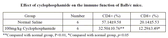
The immune inhibition effect of cyclophosphamide on Balb/c mice: To assess whether the Ta1 protein expressed in INVSc1 yeast host strain containing pYES2- Ta1 plasmid has bioactitivity effect, immune inhibition mice model was used. CD4+ and CD8+ lymphocytes in cyclophosphamide treated Balb/c mice were significantly lower than those in normal ones (32.50±10.76 vs 57.14±9.58, p<0.01, and 12.29±3.49 vs 20.14±5.53, p<0.05), which showed that the cellular immune function was significantly inhibited. The results showed that immune inhibited mice model had been constructed successfully (Table 1).
TABLE 1.
Effect of cyclophosphamide on the immune function of Balb/c mice.
Effect of expressed Ta1 protein on immune inhibited mice: To detect the function of expressed Ta1 protein, lyophilized INVSc1 yeast host strain containing pYES2-Ta1 plasmids were used to treat the immune inhibited mice with the quantity of 600μg to each mouse in normal saline (group A). INVSc1 yeast host strains containing empty pYES2 plasmid were used as negative control with the same concentration (group C), and synthesized Ta1 peptides were used as positive control with the quantity of 6μg to each mouse in normal saline (group B). As showed in Table 2, compared with group C, both group A and B significantly improved the CD8+ lymphocytes (22.74±1.09 and 18.77±4.72 vs 7.49±2.14 p<0.01), while both group A and group B had little effect on the CD4+ lymphocytes (61.86±6.94 and 65.91±4.78 vs 57.93±10.40, p>0.05).
TABLE 2.
Effect of Ta1 on immune function of Balb/c mice.

Disscussion
Ta1 plays a lot of vital roles, such as immune regulation, promoting of NK cell activity, enhancing of the anti-infectious ability of the host, promoting of viral clearance, anti-oxidant activity, inhibiting of the growth of cancer cells, etc. (Zavaglia et al., 2000). It was used widely in the clinical treatment of hepatitis B, hepatitis C, HIV and some tumors, such as melanoma, lung cancer, leukemia, squamous epithelial cancer, colon cancer, etc. (Garaci et al., 2000; Andreone et al., 2001).
There are two kinds of thymus products in clinical practice. One is an artificial chemical synthesized injection. Although it has been approved to have good effect in clinical practice, it is hard to popularize its use for its high price and the inconvenience of every day's injection. It costs about $8,000 by patient in one period of treatment in China. The other thymus product is a kind of thymosin mixture, which was extracted from animal thymus. It has a poorer treatment effect for its lower effective component (only 0.56%~1.00%). It is mingled with animal proteins and easily induces allergy in patients. It is vital to construct a new system, which can produce Ta1 by gene engineering with merits of low cost and high quality.
The results of this study showed that a Ta1 yeast expression system has been constructed successfully. The results of PCR assay, restriction enzyme assay and sequencing assay showed that pYSE2-Ta1 we constructed has correct sequence according with original one which was reported in GenBank (data not shown), and the results of Western blot assay showed that the expressed Ta1 protein can act with the Ta1 antibody.
Yeast strains are proved to be suitable for gene engineering. Yeast can be used as a kind of vector to introduce foreign genes. Whole recombinant yeast heterologously expressing mammalian mutant Ras proteins was used to immunize mice in a carcinogen-induced lung tumor model. Therapeutic immunization with the whole recombinant yeast caused complete regression of established Ras mutation-bearing lung tumors in a dosedependent, antigen-specific manner (Lu et al., 2004).
It had been identified that Saccharomyces cerevisiae can be administrated orally. Yeast strains of the species Saccharomyces cerevisiae is currently in use in the production of consumable alcohols, such as beer (Barnett 1997). Jung et al. found out that after being administered Saccharomyces cerevisiae beta-glucan orally (50 mg/day/pig) for 3 days before swine influenza virus (SIV) infection, the microscopic lung lesions of pigs induced by SIV infection were significantly minor than those of control (p < 0.05). The concentrations of IFN- γ and nitric oxide in bronchoalveolar lavage fluid from these pigs were significantly higher than those of control. These findings support that the Saccharomyces cerevisiae beta-glucan can improve the immune function when is orally administered (Jung et al., 2004).
Blanquet et al. demonstrated that engineered yeasts secreting compounds can be received directly in the digestive tract. The oral administration of the survival rate and the ability of two recombinant Saccharomyces cerevisiae strains (WppV(5)H(6) and WppGSTV(5)H(6)) to initiate the synthesis and secrete either a model peptide (peptide-V(5)H(6), MW: 5.6 kDa) or a model protein (glutathione-S-transferase -V(5) H(6), MW: 31.5 kDa) were studied in a gastric-small intestinal system simulating human digestive conditions. The WppV(5)H(6) and WppGSTV(5)H(6) strains respectively showed 83.1%+/-9.6 (n=3) and 95.3%+/-22.7 (n=4) survival rates in the model upper digestive tract after 270 min of digestion. The secretion products were detected as early as 90min after the yeast intake/gene induction in each compartment of the in vitro system, but mostly in the jejunum and ileum. The GST-V(5)H(6) concentrations in the digestive medium reached 15 ng/ml, close to values measured in batch cultures. These results open up new opportunities for the set up of drug delivery systems based on engineered yeasts secreting compounds directly in the digestive tract (Blanquet et al., 2004).
To assess whether INVSc1 yeast host strain containing pYES2-Ta1 plasmids we obtained in present work can affect the immune function with oral administration, immune inhibited mice model was used. Cyclophosphamide is a DNA alkylating agent with striking immunomodulatory properties that are widely exploited in adoptive immunotherapy regimens (Pelaez et al., 2001). Cyclophosphamide treatment is known to induce an immunosuppressed condition. Immunologic effect cells, such as B lymphocytes, T lymphocytes, and natural killer cells, are extremely sensitive to the cytotoxic properties of cyclophosphamide. Cyclophosphamide has also been used by several groups as an immunomodulatory agent against murine and human tumors (Saxton et al., 1997; Schiavoni et al., 2000). In the present study, the results showed that CD4+ and CD8+ lymphocytes in those cyclophosphamide treated Balb/c mice were significantly lower than those in normal ones (32.50±10.76 vs 57.14±9.58, p<0.01, and 12.29±3.49 vs 20.14±5.53, p<0.05). Ta1 expressed by INVSc1 yeast host strain containing pYES2-Ta1 plasmids can improve the CD8+ cell level which was suppressed by cyclophosphamide in advance, the effect coincided with the synthesized Ta1(22.74±1.09 and 18.77±4.72 vs 7.49±2.14, p<0.01). Interestingly, there was little effect of Ta1 on the CD4+ lymphocytes. The level of CD4+ lymphocytes was similar to normal control, no matter Ta1 expressed by INVSc1 yeast host strain containing pYES2-Ta1 plasmids, or synthesized Ta1 (61.86±6.94 and 65.91±4.78 vs 57.93±10.40, p>0.05). Considering that CD4+ lymphocytes almost reach normal levels at 15th day (57.93±10.40), it is reasonable there was little change in CD4+ lymphocyte level. The reason why CD4+ lymphocytes will return back to normal values is still unknown, it is likely to relate with the cease of cyclophosphamide, which is worth further investigation.
In conclusion, a high effective yeast expression system for Ta1, that is INVSc1 yeast host strain containing pYES2-Ta1 plasmids, was constructed successfully, and the Ta1 expressed by this system can improve the level of CD8+ cells in Balb/c mice treated with cyclophosphamide in advance.
Acknowledgements
This project was funded from Natural Science Foundation of Zhejiang Province (No. ZD9904) and the key project from Science and Technology Department of Zhejiang Province (2003C13015).
References
1. Ancell CD, Phipps J, Young L (2001). Thymosin alpha-1. Am J Health Syst Pharm 58: 879-885. [ Links ]
2. Andreone P, Cursaro C, Gramenzi A, Margotti M, Ferri E, Talarico S, Biselli M, Felline F, Tuthill C, Martins E, Gasbarrini G, Bernardi M (2001). In vitro effect of thymosin-alpha1 and interferon-alpha on Th1 and Th2 cytokine synthesis in patients with chronic hepatitis C. J Viral Hepat 8: 194-201. [ Links ]
3. Barnett JA (1997). A historical survey of the study of yeasts. In: Yeast Sugar Metabolism. Zimmermann FK and Entian K-D, Eds. Technomic Publishing, Lancaster, pp. 1-33. [ Links ]
4. Billich A (2002). Thymosin alpha1. SciClone Pharmaceuticals. Curr Opin Investig Drugs 3: 698-707. [ Links ]
5. Blanquet S, Antonelli R, Laforet L, Denis S, Marol-Bonnin S, Alric M (2004). Living recombinant Saccharomyces cerevisiae secreting proteins or peptides as a new drug delivery system in the gut. J Biotechnol 110: 37-49. [ Links ]
6. Chien RN, Liaw YF, Chen TC, Yeh CT, Sheen IS (1998). Efficacy of thymosin a1 in patients with chronic hepatitis B: A randomized, controlled trial. Hepatology 27: 1383-1387. [ Links ]
7. Garaci E, Pica F, Rasi G, Favalli C (2000). Thymosin alpha 1 in the treatment of cancer: from basic research to clinical application. Int J Immunopharmacol 22: 1067-1076. [ Links ]
8. Hannappel E, Huff T (2003). The thymosins£¬prothymosin alpha, parathymosin, and beta-thymosins: structure and function. Vitam Horm 66: 257-296. [ Links ]
9. Jung K, Ha Y, Ha SK, Han DU, Kim DW, Moon WK, Chae C (2004). Antiviral effect of Saccharomyces cerevisiae beta-glucan to swine influenza virus by increased production of interferongamma and nitric oxide. J Vet Med B Infect Dis Vet Public Health 51: 72-76. [ Links ]
10.Kullavanuaya P, Treeprasertsuk S, Thong-Ngam D, Chaermthai K, Gonlachanvit S, Suwanagool P (2001). The combined treatment of interferon alpha-2a and thymosin alpha 1 for chronic hepatitis C: the 48 weeks end of treatment results. J Med Assoc Thai 84 Suppl 1: S462-468. [ Links ]
11. Lu Y, Bellgrau D, Dwyer-Nield LD, Malkinson AM, Duke RC, Rodell TC, Franzusoff A (2004). Mutation-selective tumor remission with Ras-targeted, whole yeast-based immunotherapy. Cancer Res 64: 5084-5088. [ Links ]
12. Moody TW, Tuthill C, Badamchian M, Goldstein AL (2002). Thymosin alpha1 inhibits mammary carcinogenesis in Fisher rats. Peptides 23: 1011-1014. [ Links ]
13. Mutchnick MG, Appelman HD, Chung HT, Aragona E, Gupta TP, Cummings GD, Waggoner JG (1991). Thymosin treatment of chronic hepatitis B: A placebo-controlled pilot trial. Hepatology 14: 409-415. [ Links ]
14. Pelaez B, Campillo JA, Lopez-Asenjo JA, Subiza JL (2001). Cyclophosphamide induces the development of early myeloid cells suppressing tumor cell growth by a nitric oxide-dependent mechanism. J Immunol 166: 6608-6615. [ Links ]
15. Rasi G, Pierimarchi P, Sinibaldi Vallebona P, Colella F, Garaci E (2003). Combination therapy in the treatment of chronic viral hepatitis and prevention of hepatocellular carcinoma. Int Immunopharmacol 3: 1169-1176. [ Links ]
16. Saruc M, Ozden N, Yuceyar H (2003). Thymosin in the treatment of HBeAg-negative chronic hepatitis B. Med Sci Monit 9: RA198-202. [ Links ]
17. Saxton ML, Longo DL, Wetzel HE, Tribble H, Alvord WG, Kwak LW, Leonard AS, Ullmann CD, Curti BD, Ochoa AC (1997). Adoptive transfer of anti-CD-activated CD4+ T-cells plus cyclophosphamide and liposome-encapsulated interleukin-2 cure murine MC-38 and 3LL tumors and establish tumor-specific immunity. Blood 89: 2529-2536. [ Links ]
18. Schiavoni G, Mattei F, Di Pucchio T, Santini SM, Bracci L, Belardelli F, Proietti E (2000). Cylophosphamide induces type I interferon and augments the number of CD44 (hi) T lymphocytes in mice: implications for strategies of chemoimmunotherapy of cancer. Blood 95: 2024-2030. [ Links ]
19. Zavaglia C, Airoldi A, Pinzello G (2000). Antiviral therapy of HBVand HCV-induced liver cirrhosis. J Clin Gastroenterol 30:234- 241. [ Links ]
Received on October 10, 2004.
Accepted on May 23, 2005.

