


 Inglés (pdf)
 Articulo en XML
 Referencias del artículo
 Enviar articulo por email
 Citado por SciELO
  Similares en
SciELO
Similares en
SciELO  uBio
uBio 
 Permalink
Introduction
The onion (Allium cepa L. var. cepa) is one of the most cultivated horticultural species to produce bulbs (Galmarini, 2018). According to FAO, world production in 2021 was 151 million tons, covering approximately 6 million ha; in Argentina, 16,000 ha of onions were cultivated in 2021 with a production of 600,000 tons (FAOSTAT, 2021). The implantation of the crop can be done by sowing seed or planting bulbs. Argentina not only stands out for the production of bulbs but also for seed production, the most important areas are located in central-western provinces of San Juan and Mendoza. Fertile or open-pollinated cultivars and also first-generation hybrids (F1), produced by crossing pure or partially homozygous lines with a male-sterile line (MSL), are used for seed production. Male sterility was discovered in onion in 1925 (Saini & Davis, 1969) and since then it has been a valuable resource for hybrid seed production (Havey, 2000; Kamenetsky & Rabinowitch, 2002; Kik, 2002; Engelke et al., 2002, 2003). Onion hybrid seeds have been extensively produced all over the world using cytoplasmic-genic male sterility (CGMS) based systems. There are two main sources of cytoplasmic male-sterility identified as S and T, which have been genetically characterized. S type results from the interaction of a cytoplasmic factor S and a single nuclear restorer gene Ms (Colombo & Galmarini, 2017, and references therein). The system of CGMS is defined as the inability to produce viable pollen grain in plants. The lower average seed yield in F1 hybrids may be the result of deficient pollination and factors related to flower morphology or nectar composition of the MSL (Parker, 1982; Céspedes et al., 2004; Soto et al., 2013, 2015; Gatica Hernández et al., 2019).
Onion flowers are grouped in umbels, which have between 50 and 2,000 flowers (Fig 1A-B; Brewster, 2001; Kavitha & Reddy, 2018). Flowers are protandric: in the fertile variety the anthers mature normally and pollen is released before the stigma becomes receptive (Currah & Ockendon, 1978). The flower bears a perigonium consisting of two whorls of three tepals; the androecium is composed of two whorls of three stamens, and the gynoecium is composed of three carpels fused in a superior ovary, with axillary placentation and two ovules per locule (Gaviola, 2007). In plants that are good seed producers, there are usually three to four mature seeds in each fruit. In male-sterile plants, dehiscence of the anthers fails, and the pollen production is aborted (Holford et al., 1991).
Offering rewards such as nectar is one of the plant’s resources for attracting pollinators (Fahn, 1979; Bernardello, 2007; Pacini & Nepi, 2007; Nepi et al., 2012). There are numerous studies on various species that show that the size, shape or color of different floral parts are related to pollinator visits (López & Galetto, 2002; Pernal & Currie, 2002; Silva et al., 2003;Abrol, 2005; Poveda et al., 2005; Ford & Johnson, 2008; Human & Nicolson, 2008; Steenhuisen et al., 2010). The flowers of many monocotyledons present gynopleural floral nectaries (GN), also called septal nectaries (Van Heel, 1988; Smets et al, 2000). The GN are nectar-secreting cavities consisting of the incomplete fusion of the carpel walls in the septal region (Smets & Cresens, 1988; Pacini & Nepi, 2007; Odintsova et al., 2013; Fishchuk & Odintsova, 2020). Fritsch (1992) compared the position of the GN of 160 species of Allium, including A. cepa, highlighting the importance of this characteristic in the confusing taxonomy of the genus Allium (Gurushidze et al., 2007). Contrary to the scant recognition of structural studies on floral nectaries of onion, there is a profuse bibliography of nectar chemisüy and the relationship with pollinators (Syamasundar & Panchaksharappa, 1976; Kumar & Gupta, 1993; Silva et al., 2000, 2004; Soto et al., 2015; Gillespie et al, 2015, Divija et al., 2022).
In many cases, the structure of the flowers is organized in such a way that the place of nectar production is different from the place of its presentation, i.e., where the nectar accumulates and is offered to the visitors, which is called secondary presentation (Fahn, 1979; Vogel, 1998; Pacini & Nepi, 2007). Kavitha & Reddy (2018) described Allium flowers as bowl-shaped with the nectary hidden in the lower part of the ovary. The nectar produced accumulates in the cup located between the base of the ovary and the inner whorl of stamens, which have staminal filaments with a widened base (Fig. 1B). According to Brewster (2001), this area is easily accessible to many types of pollinating insects. It has been demonstrated that there is variability between the fertile and male-sterile onion lines regarding the preference of bees to visit their flowers (Soto et al., 2013, 2015, 2021).
The aim of the present study is to extend the morpho-anatomical descriptions of Allium cepa flowers, to re-evaluate the three-dimensional structure of the gynopleural nectary in relation to the floral parts and to explain the phenomenon of secondary nectar presentation. It is also intended to determine whether there are differences in the structure of the nectary in fertile cultivars and male-sterile lines. In addition, the floral anatomy will be studied in depth with emphasis on the anthers of the fertile and male-sterile lines to understand the availability of pollen in the varieties cultivated in Argentina.
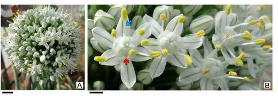
Fig. 1: Allium cepa. A: Inflorescence of fertile variety. B: Flowers, the red arrow indicates the inner stamen with a broad base and the blue arrow to the outer stamen with a thin staminal filament. Scales= A: 0.5 cm, B: 0.1 cm.
Materials and Methods
Four onion accessions were used, one male-fertile variety (FV, Valcatorce INTA; Galmarini, 2000) provided by INTA EEA La Consulta and three male-sterile lines (MSL, ON.192A, ON.013A, and ON.108A) provided by the Enza Zaden Seed Company. Vouchers (fixed material) were deposited at CTES herbarium, voucher number: Valcatorce INTA AMG N° 528, ON.192A AMG N° 529, ON.013A AMG N° 530, and ON.108A AMG N° 530.
Flowers of A. cepa were fixed in FAA (formaldehyde, alcohol 70%, acetic acid, 90:5:5). The material was dehydrated through a tertiary butanol series and embedded in paraffin (Gonzalez & Cristóbal, 1997; Ruzin, 1999), microtomized using a Microm HM350 rotary microtome in 12 pm transversal (TS) and longitudinal sections (LS), and finally stained in safranin and astra blue combinations (Luque et al., 1996). A Leica MZ6 light microscope (LM) with a digital imaging system was used for the observations.
For scanning electron microscopy (SEM), FAA-fixed material was dehydrated in an increasing acetone series, critical-point dried using liquid CO2 and sputter coated with gold-palladium. The gold-coated samples were photographed with a SEM Jeol LV5800 (JEOL Ltd., Tokyo, Japan), at 20 Kv in the Service of Electron Microscopy facility at Universidad Nacional del Nordeste (Corrientes, Argentina).
Results
Floral morphology and anatomy
The morphological structure of fertile flowers and male-sterile lines is the same, the difference lies in the pollen production. The flowers of A. cepa have two whorls of free tepals, three outer and three inner, white with a green central line on both sides (Fig. 1B).
The androecium consists of two whorls of three stamens each, those of the outer whorl have slender staminal filaments. The three inner stamens have tricuspidate staminal filaments, with basally widened tepaloid expansions finishing in two free ends and a third that lengthens and supports the anther (Figs. 1B; 2A-C). The widened bases of the inner staminal filaments form a cup-shaped area where the floral nectar accumulates. The anthers are bithecal, versatile, and dorsifixed (Fig. 2A-C). In both FV and MSL flowers, a dehiscence line is visible externally on the anthers, but thecae of the MSL do not open or release pollen grains (Fig. 2B-C).
Both FV and MSL present the same anatomy in the anther wall and the stomium zone, the differences between them are due to the process of microgametogenesis (Fig. 2D-M).
In mature anthers of FV, the wall of the thecae is only composed of an exothecium and endothecium (Fig. 2 D-F). The exothecium bears cells with thickened cellulosic walls which are covered with a finely reticulated cuticle, which lacks stomata (Fig. 2G). The endothecium is composed of a single layer of thin-walled cells with U-shaped lignified thickenings; the bars of these thickenings are free, covering the inner tangential wall and the side walls without reaching the outer tangential wall (Fig. 2H). The stomium zone in FV has small epidermal and endothecial cells and longitudinal dehiscence occurs through these cells (Fig. 2F). In the ripe anther of FV, orbicules are observed covering the inner walls of the pollen sacs. Pollen is monosulcate and oblate in shape, with a rugulate sculpture (Fig. 2I).
The anthers of FV have the same organization in the anther wall and stomium zone (Fig. 2J-L) Despite having an endothecium with well-developed thickenings, there is no dehiscence. The stomium also has small-sized cells. The difference with fertile anthers lies in the volume of the pollen sacs. The sacs have collapsed and there is cellular debris inside. In MSL the anthers do not produce pollen; the remains of sporogenous cells or aborted microspores can be observed, agglutinated by tapetal cellular residues (Fig. 2M). In the anthers of MSL, the cells of the stomium zone, endothecium, and epidermis are not affected, they have the same cellular conformation as in FV; however, no dehiscence occurs in these anthers.
The tepals and tepaloid bases of the staminal filaments (in both FV and MSL) have the same anatomical structure: a compact parenchyma, covered by a unistratified epidermis, without stomata (Fig. 2J). The filiform portion of all the staminal filaments is thin; the colorless and compact parenchyma is covered by a unistratified epidermis of cells arranged in palisade, without stomata, covered with a finely striated cuticle (Fig. 2D, J).
Gynoecium and Nectary
There is no difference in the structural or vascular organization between FV and MSL flowers. The ovary is superior, tricarpellar, trigonous, and slightly flattened. The carpels are organized in an axillary placentation with two ovules per locule (Figs. 3C, 4). The style is single and gynobasic, it is located in an invagination of the apical portion of the ovary; the stigma is an inconspicuous knob that completes its elongation after the dehiscence of the anthers occurs (only in FV).
The complex organization of the nectary can only be understood by analyzing the exomorphology of the gynoecium and its anatomy through a sequence of transversal and longitudinal serial sections of the ovary (Figs. 3-4).
Externally the ovary has three grooves that protrude at the edges, which accompany the dorsal bundle of each carpel along its path (Figs. 3A-F, 4J-L). The carpel wall in the dorsal bundle area presents an external groove, which is the only area of the epidermis with stomata that lack a substomatal chamber.
The lateral sides of the ovary are flat and are constituted by two adjacent carpels, without delimitation of the suture line (Fig. 3B-D). By removing the ovary and observing it from the base, three elongated and pocket-shaped openings are visible; they consist of crests of carpellary tissue that cover the ovary laterally, delimiting three cavities or néctar pockets (Fig. 3C-E). Each crest corresponds to two contiguous carpels (Fig. 3C).
In cross-sections of the ovary it is observed that the portion of two adjacent carpels folds inwards forming the septum and delimiting the axillary placentation (Figs. 3D-E; J-L). In the septum, the suture between the walls of the carpels is incomplete and forms three grooves, each corresponding to GN or septal nectaries (Figs. 3D-E; H-K). In this area, the epidermal cells are arranged in a palisade (Fig. 3F-G). The epidermis and 1-2 layers of subepidermal parenchyma of the carpel are differentiated into nectar-secreting tissue, characterized by a very dense cytoplasm with a central nucleus and no perceptible vacuoles; the walls are cellulosic and thin (Fig. 3F-G).
The rest of the septum, as well as the carpel wall, is formed by a compact fundamental parenchyma. Inside the pocket, the crest has an inner longitudinal groove (Fig. 3E). Each crest over the GN consists of 3-4 layers of ground parenchyma, covered by a unistratified epidermis, without stomata; there are no secretory cells and they lack vascularization (Fig. 3H-J).
In a radial longitudinal section passing through the septum and nectary, it can be seen how the apical portion of GN is connected to the nectariferous pocket (Fig. 3H-J). The nectar produced and excreted into the septum area thus finds a place to exit into the nectariferous pocket and accumulates between the ovary and the tepaloid base of the staminal filaments.
Each locule bears two ovules, inserted in the axils of the carpels, in its basal portion (Fig. 3A, D, H, K). The ovules are campylotropous with curved nucellus (Fig. 3K-L). Both integumento have between 6-8 layers of thickness, which are multiplied in the micropyle. The external epidermis of the inner integument presents cells arranged in a short palisade. The nucellus is inconspicuous, only one layer of cells separates the embryo sac from the endostome. The chalaza has small cells of dense cytoplasm, with the appearance of hypostasis; this tissue even reaches the vascular bundle. The epidermis of the placenta is differentiated in secretory tissue, constituting a placental obturator (Fig. 3L).

Fig. 2: Flowers of Allium cepa. A-C, J-M: SEM; D-I: LM. A-B: Longitudinal section of fertile variety (FV) flower. C: Androecium and tepals in male-sterile line (MSL) flowers; asterisks in B-C indicate tricuspidate tepaloid expansions of staminal filaments. D-I: FV anther. D: Transversal section of almost dehiscing anther. E: Detail of anther wall showing endothecial cells with fibrous thickenings and pollen grains. F: Stomium zone. G: Superficial view of anther epidermis. H: Paradermal section showing endothecium with fibrous thickenings. I: Pollen and inner face of tapetal membranes. J-M: MSL anther. J: Tepals and anther with collapsed pollen sacs. K: Detail of anther and collapsed pollen sac. L: Indehiscent stomium zone. M: Abortive microspores inside MSL anther. Abbreviations: ct: collapsed tissue, et: exothecium, is: inner stamen, it: inner tepal, np: nectary pocket, nt: endothecium, os: outer stamen, ot: outer tepal, ov: ovule, ova: ovary, s: stomium, sf: staminal filament, st: style. Scales= A-C: 500 pm; D, J: 200 pm; E-F, K-L: 50 pm; G-H: 20 pm; I, M: 10 pm.

Fig. 3: Gynoecium and gynopleural nectary of Allium cepa. A-F, I, K: SEM; G, H, J, L: LM. A: Longitudinal section of fertile variety (FV) flower; the asterisk shows the aperture for néctar exit. B: Apical view of ovary. C: Basal view of ovary showing the nectary pocket apertures; white lines indícate the limits between carpels, white arrowpoint indicate dorsal slots (in B-D). D: Transversal section (TS) of ovary. E-F: False color in SEM images: pink = cytoplasm, blue = nucleus. E: Detail of the ovary in TS showing the gynopleural nectary (GN: pink cells) and nectary pocket covered by fíat crest. F-G: Glandular epidermis of GN. H: Longitudinal section of male-sterile line (MSL) flower indicating an ovule in the left lobe and the nectary to the right, covered by a crest (arrow), the nectar accumulation area between ovary and anther is indicated by an asterisk. I: Nectary pocket and crest in detail. J: Detail of H showing GN; arrow indicates the apical opening for nectar exit to the nectary pocket (asterisk). K-L: Ovule. Abbreviations: cr: crest, gn: gynopleural nectary, it: inner integument, np: nectary pocket, ob: obturator, ot: outer integument, ov: ovule, st: style. Scales= A-D: 500 pm; E, H, J: 200 pm; I, K-L: 100 pm; F-G: 20 pm.
In the longitudinal section of the gynoecium, the style is inserted in the center of the ovary, almost at the base, due to the invagination of the carpellaiy wall (Fig. 3A, H). The style is hollow, the central channel is covered with cells whose radial walls are loosely arranged; the entire inner part of this epithelium and the spaces between the walls are filled with mucilage. The stigma is lined by cells with convex outer walls and, consequently, has a papillose surface.
Floral vascularization (Fig. 4): the flower peduncle is innervated by 6-9 vascular bundles, which are united forming a hexagonal ring in the receptacle (Fig. 4A-D). Six traces are detached towards the periphery, each one is divided periclinally, the six external traces vascularize the tepals and the six internal ones innervate the short staminal tube (Fig. 4E-F). Each stamen remains vascularized by a small bundle that runs through the staminal filament to the connective tissue between the anthers. The tepaloid projections of the staminal filaments of the inner stamens lack vascularization other than that of the filaments (Fig. 4G). From the central vascular ring, three traces separate and become the dorsal bundles of each carpel (Fig. 4F-H). The vascular remnant adopts a triangular shape dividing into ventral and placental bundles that will innerve the ovules (Fig. 4G-L). The style lacks a vascular supply.

Fig. 4: Allium cepa floral vascularization and gynopleural nectary (GN) position. A: Longitudinal section of ovary showing different levels of transverse sections in diagrams B-L: (glandular tissue shaded, vascular bundle in black) and transversal sections (M-R). A-D: Peduncle region. E-F: Receptacle. G-J: Ovary and staminal filaments, tepals were not drawn. K-L: Ovary. M-R: LM of gynopleural nectary transection; nectary pocket is indicated with asterisk. Abbreviations: cr: crest, cs: nectar accumulation area, dc: dorsal carpellary vascular bundle, fis: broad base of inner staminal filament, fos: thin outer staminal filament, gn: gynopleural nectary, it: inner tepal, ot: outer tepal, np: nectary pocket, ov: ovule, ova: ovary, st: style. Scales= A-L: 1 mm; M-R: 0.5 mm.
In the series of sections in Fig. 4A, M-R, the relative positions of the gynopleural nectary and the nectariferous pocket can be observed.
Discussion and ConclusionsThe anatomical study of the flower showed that there are no differences between the male-fertile and male-sterile lines involving the floral structure in general or the gynoecium or nectary structure in particular. The fundamental difference between FV and MSL lines is the interruption of male gametogenesis and the process of anther dehiscence. The timing of abortion is concentrated in the late stages of microsporogenesis when the callose dissolves and microspores are released into the anther locules. In the present study, the presence of collapsed microspores was observed among cellular remains presumably of tapetal origin. This is consistent with previous embryological and anatomical studies in which it was observed that, although meiosis occurred normally, male sterility is produced by hypertrophy of the tapetal cells and/or pollen degeneration (in onion: Monosmith, 1928; Saini & Davis, 1969; Tchórzewska et al., 2017; in garlic: Shemesh-Mayer et al., 2015; in chives: Engelke et al., 2002). The wall structure of fertile and sterile anthers was not analyzed in the aforementioned studies. In onion, the programmed cell death of the sexual cells does not affect the development of the anther wall, septum and stomium zone, which present a normal structure. With respect to anther dehiscence, the MSLs show anthers in which a dehiscence line is recognized externally; anatomically, this region is the stomium, structurally typical with smaller cells. The endothecium of the anthers in MSL forms thickenings identical to that of the FV. However, despite this normal anther wall, dehiscence of the locules does not occur.
Anther dehiscence is a process that involves changes in the sterile tissues (endothecium and development of ligno-cellulosic thickening, septum, and stomium defined as weakened regions susceptible to lysis and rupture, general dehydration of the anther) and sexual cells (mature pollen swelling). This process has been widely studied by several authors (Keijzer, 1987, 1999; Bonner & Dickinson, 1989; Scott et al., 2004; Wilson et al., 2011). In Allium cepa a great effort has been put into genetic and molecular studies to understand the process of cytoplasmic male sterility and its contributions to future breeding programs (Ahmad et al., 2020; Manjunathagowda et al., 2021; Yu & Kim, 2021). However, none of these studies took into account the development of the exothecium or the cells of the stomium zone. Only García et al. (2006) studied the mechanism of anther opening, but in Allium triquetrum L, and they associated it with the variation in moisture content and consequent dehydration, considering that dehiscence is a phenomenon temporarily independent of the opening of the stomium. Our study, which included fertile and male-sterile lines, shows that, as both types of flowers have the same anther wall and stomium structures, it is the swelling of pollen in the later stages of maturation and the pressure exerted on the thecal walls that contribute to the dehiscence of fertile anthers. The absence of mature pollen in the MSL prevents dehiscence of the anthers, despite having a structurally normal stomium and endothecium. This anther opening mechanism is the same as that described for rice by Matsui et al. (1999).
Studies carried out on floral exomorphology that influence seed production of hybrid onions, showed that there was a high and consistent correlation among some floral morphometric characters, bee visits and seed production; the most interesting being the surface of the external anther and the length of the style (Soto et al., 2018). The length of the style is the morphometric character that allows differentiation among open-pollinated and male-sterile plants, as well as between different MSLs in their ability to attract bees. The length of the inner tepals presented a negative and highly significant correlation with the length of the style; the former floral character is easier to measure than the length of the style and could facilitate the selection of lines with greater potential for seed production (Maldonado, 2014; Soto et al., 2018).
Gynopleural nectaries
Unfortunately, nectaries have not received much attention in studies of onions. Jones & Emsweller (1936) studied the ontogenetic development of flowers, focusing on the macrogametophyte, but they did not pay attention to the nectaries, despite their excellent drawings showing the ovary and the position of the ovules in three dimensions.
Septal nectaries, correctly called gynopleurals (Smets & Cresens, 1988), are present in most monocotyledon orders (except Liliales) and thus in many families, such as Amaryllidaceae, Asparagaceae, Asphodelaceae, and Iridaceae, among others (Weberling, 1992; Vogel, 1998; Smets et al., 2000; Rudall, 2002; Bernardello, 2007; Odintsova et al., 2013: Fishchuk & Odintsova, 2020, 2021).
In the genus Allium, there is variation in the form of the gynoecium and also in the presence or lack of appendages or ovarian processes (Fritsch, 2001; Choi et al., 2011, 2012). These processes may be either absent (naked ovary) or present, in which case they can be of two types: apical crest-like (characteristic of northern North American species) or basal hood-like processes (in northeastern Asian species, subgenus Cepa, in which A. cepa is included). Choi et al. (2011, 2012) stated that the septal nectary opening and development of ovarian processes have taxonomic importance; however, despite the importance of this feature it has not received attention in other structural and anatomical studies.
In particular, in A. cepa the structure of GN was first analyzed by Syamasundar & Panchaksharappa (1976). Based on nectary histochemistry, these authors only mentioned the existence of three septa with nectariferous tissue. Heel (1988) analyzed the ontogeny of the pistil in several monocotyledons with septal nectaries, including Allium fistulosum L. In this species, the GN opening is narrow and only covered by a small ovarian process (called an epidermal flap), which formed late in the ontogeny of the gynoecium. Allium fistulosum does not develop a crest and pocket like A. cepa.
Fritsch (1992) analyzed the shape, position, and excretory canals of GN in 160 species of Allium, highlighting the importance of this character in taxonomic research. This author mentioned that in sect. Cepa (in which A. cepa is included) “the canal consists of an inner, tube-like part, which opens from the radial outer side into a pocket-like, tangentially widened outer part ending in the lower third of the ovary”, this being a unique feature in the genus Allium. Unfortunately, he showed this in a diagram of the longitudinal section of the ovary, in which it is difficult to understand the actual three-dimensional structure of this description. The same type of descriptions and drawings can be found in Di Fulvio (1973). Recently, Fishchuk (2022) published the vascularization of the flowers of A. cepa and established three vertical zones in the gynoecium: 1) a synascidiate zone at the locule base, 2) a symplicate structural zone, which contains the ovules, and 3) a hemisymplicate zone, which occupies the upper part of the locule; our results agree with these observations in both FV and MSL.
The onion flower has a complex organization and the production, secretion, and presentation of nectar are organized in three well-defined regions:
Nectar-producing zone: located in the three interlocular septa, radially arranged and separating the locules of the ovary. The glandular tissue is composed of the epidermis and subdermal parenchyma, in the area formed by the incomplete suture line between carpels. This area is the site of nectar secretion and the septal (GN) nectary itself. The nectar outlet is in the apical zone and is hidden by the flat crest.
Nectar-discharge zone: constituted by the three nectary pockets, covered by a flat crest consisting of the walls of adjacent carpels that cover the lateral walls of the ovary. The interior of this pocket - both the ridge tissue and the walls of the ovary - lack nectar-secreting tissue. Each pocket connects to the production area in a short section located at the top of the secretory area. This second zone opens to the outside at the base of the ovary, through three large openings or pockets.
Accumulation and harvest zone: nectar produced in the GN and expelled through the apical opening passes through pockets and accumulates in a third collecting zone. These areas are composed by the widened base of the three inner stamens, opposed to the aperture of the nectaries. In this area, the excreted nectar can be collected by insects that visit the flower.
The vertical zoning of the GN was described in the flowers of Dracaena fragrans (L.) Ker Gawl., Sansevieria parva N.E. Br. and S. trifasciata Prain (Asparagaceae) by Odintsova et al. (2013). These authors also recognize three zones: 1) the distinct nectary formed by three cavities at the base of the ovary, 2) the zone of common nectary in the ovary center where the cavities join, and 3) the external nectary zone, that corresponds to the upper part of the ovary where the septal grooves fuse with the nectariferous cavities and nectary splits opened to the exterior. Unlike A. cepa, in these species there is no accumulation area and harvest zone, since the GN of Dracaena and Sansevieria species lack crests and pockets. Onion floral nectaries are a clear example of secondary presentation of nectar, it is produced in one place (in the ovarían septa) and offered in a different place (cavities between broad staminal filaments and the ovary). In the onion, there is also another area dedicated to the transport between the place of production and the accumulation and harvesting area. A point that would be interesting for further research, by means of an ontogenetic study, is the origin of the crests that cover the nectary pockets. The correlation found in some floral and chemical traits with onion seed yield reported in other studies may be a great contribution for onion breeders, who could select lines that have promising traits for pollinator attraction and, consequently, seed yields. Further studies are also required concerning the heritability of floral traits.
Acknowledgments
To INTA EEA La Consulta (Mendoza, Argentina) and Enza Zaden Seed Company for supplying us with the botanical material. This research was partly supported by the Universidad Nacional del Nordeste to AMG (Grants N° 16A003 and 20P001, SGCyT-UNNE).
Author Contributions
AMG: performed the slides, interpreted the anatomic analysis, made up the figures and wrote the manuscript. IM, CG & IP: collected field material and reviewed the text.
Primary Data Availability
The primary research data are available in CONICET's institutional repository: https://ri.conicet.gov.ar/handle/n336/193050.

