


Servicios Personalizados
Revista

Articulo
 Inglés (pdf)
 Articulo en XML
 Referencias del artículo
 Enviar articulo por email
Indicadores
-
 Citado por SciELO
Links relacionados
-
 Similares en
SciELO
Similares en
SciELO  uBio
uBio
Compartir

 Permalink
Phyton (Buenos Aires)
versión On-line ISSN 1851-5657
Phyton (B. Aires) vol.83 no.2 Vicente López dic. 2014
ARTÍCULOS ORIGINALES
Delimitation of Azadirachta indica A. Juss. from Melia azedarach L. (Meliaceae Juss.) based on leaf morphology
Reconocimiento de Azadirachta indica A. Juss. a partir de Melia azedarach L. (Meliaceae Juss.) en base a la morfología foliar
Usama K Abdel-Hameed
Ain Shams University, Faculty of Science, Botany Department, Abassia, Cairo, Egypt.
Address Correspondence to e-mail: usama_abdelhameed@sci.asu.edu.eg; usa_uk@hotmail.com Tel: +20 122 3616259.
Recibido / Received 11.IX.2013.
Aceptado / Accepted 18.IX.2013.
Abstract. In Egypt there are two different species that are commercially marketed under the same trade name of Neem: one is Azadirachta indica A. Juss., and the other is Melia azedarach L. In this paper, leaf morphological characters (e.g., lamina architecture, stomatography, petiole and blade micromorphology) of both taxa were described and illustrated to aid in the identification and differentiation between the two misidentifed taxa. The obtained results concluded that leaf morpho-anatomical characters will not only provide criteria for their correct taxonomic authentication, but would also serve as future standard data for the quality assessment of the pharmaceutical preparation of botanical drugs.
Keywords: Morphology; Leaf architecture; Stomatography; Azadirachta indica; Melia azedarach; Meliaceae.
Resumen. En Egipto hay dos especies diferentes que se comercializan bajo el mismo nombre comercial: Neem. Una de las especies es Azadirachta indica A. Juss., y la otra es Melia azedarach. En este artículo se describen e ilustran características morfológicas foliares, (ej., arquitectura foliar, fotografías foliares a escala de micra, pecíolo y micromorfología de la lámina) a fin de contribuir a una correcta identificación y diferenciación entre ambas especies, incorrectamente identificadas al presente. Se concluye que las características morfoanatómicas foliares no solo proveen criterios para una adecuada clasificación taxonómica de ambas especies, sino también proveen de datos estándar futuros para una correcta evaluación de calidad en la preparación farmacéutica de drogas botánicas.
Palabras clave: Morfología; Arquitectura foliar; Fotografías foliares a escala de micras; Azadirachta indica; Melia azedarach; Meliaceae.
INTRODUCTION
The increase in the demand for herbal medicines may lead to adulteration and misidentification of the raw material (Ahmad et al., 2010). The authentic botanical identification of herbal drugs is the base for the future development of the pharmacognosy (Ahmad et al., 2008, 2009). Multiple approaches of taxonomic analysis (e.g., documentation of the biological source and morphological characters) are needed for describing herbal drugs in a systematic manner to reach authentication, and thus maintaining herbal drug efficacy (Girach et al., 1998; Sultana et al., 2011). In some herbal markets, different taxa are sold erroneously under the same common name. For example, the two different taxa Azadirachta indica and Melia azedarach are sold under the trade name of Neem, due to their morphological similarities. This leads to the misuse of Neem plants for treatment of specific diseases (Ahmad et al., 2010; Khan et al., 2011).
In the present study, taxonomic analyses allowed to distinguish the appropriate herbal drug A. indica (Neem) from the erroneously used M. azedarach (Zanzalakht). This will contribute to solve the confusion problems faced by herbalists, pharmacists, taxonomists and medicinal herb traders. Thereafter, the objective of this study was to determine leaf morphological traits which allow diferentiation between the two misidentified taxa.
The family Meliaceae includes 600 species and 52 genera among which is Azadirachta (Reveal et al., 1999). The US National Academy of Sciences published a report in 1992 entitled 'Neem - a tree for solving global problems' to express the importance of A. indica (El-Hawary et al., 2013). Since ancient times, various parts of the Neem tree have been used as a traditional medicine against various human ailments (Kraus, 1995; Singh et al., 1996). Azadirachtin is one of the chemical compounds isolated from A. indica, determined by HPLC techniques (Soni, 2012). It possesses many medicinal properties (e.g., anti-inflammatory, antioxidant, antipyretic, analgesic, immunostimulant, diuretic, hypoglycaemic, cardiovascular, antimicrobial, antiviral, antimalarial and anthelmintic activities: El-Hawary et al., 2013).
MATERIALS AND METHODS
Fresh, mature leaves of both A. indica and M. azedarach, grown in Egyptian botanical gardens, were collected and used in the present study. Identification was confirmed following Bailey, (1949), Metcalfe & Chalk (1950), Parrotta (2001) and Ross (2005).
The studied taxa were further matched against dried specimens in the Herbaria of Ain Shams University (CAIA), Cairo University (CAI), Flora & Phytotaxonomy & Agriculture Research Center (CAIM) and Orman Botanical Garden. Voucher specimens of the studied taxa were deposited in CAIA.
Macromorphological attributes of leaves were described for the investigated specimens. Lamina vein architecture investigation was made without any chemical treatment using a binocular stereo zoom light microscope (Bel Photonics). Leaves were first illuminated using glass boxes designed by the author, and then photographed by a digital camera Cannon PowerShot G12. The photographs were inverted to black and white mode for documentation. Leaf architectural terminology followed LAWG (1999).
Stomatography was carried out following the traditional method of Stace (1965). The photomicrographs were taken using a Reichert Microstar IV microscope at the Plant Taxonomy Research Laboratory, Botany Department, Faculty of Science, Ain Shams University, Cairo, Egypt. Descriptive terminology of epidermal characteristics was based on works of Metcalfe & Chalk (1950), Murley (1951), LAWG (1999) and Prabhakar (2004).
Anatomical investigations were performed after fxation and preservation of fresh materials in Formalin-Aceto-Alcohol (F.A.A). Petioles and leaves were hand-microtomed at 10-16 µ. Safranin (2%) and light green (1%) were used for double staining.
RESULTS
Leaf macromorphology. Azadirachta indica leaves have the following characteristics: alternated, crowded near the end of branches, simply pinnated, imparipinnated, 20-40 cm long, exstipulated, light green in colour, with 2 pairs of glands at the base, otherwise glabrous; petiole 2-7 cm long, subglabrous; rachis channeled above; leafets 8-19 with very short petioles, alternate proximally and more or less opposite distally, ovate to lanceolate, sometimes falcate 3.5-10 x 1.2-4 cm, glossy, margin serrated with simple tooth order, two teeth per centimeter, irregular teeth spacing, each tooth with straight apical side and concave basal one, simple tooth apex, angular sinus, apex acuminate, very oblique at the base. Melia azedarach leaves are alternated, 20-40 cm long, bipinnated or occasionally tripinnated. Leafets 3-11, with serrate margin and compound tooth order, five teeth per centimeter, regular tooth spacing, apical and basal sides convex in shape, simple tooth apex, rounded sinus shape (Fig. 1, a-b).
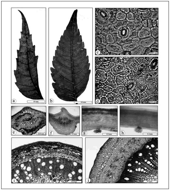
Fig. 1. Photographs illustrating foliar characters of Azadirachta indica (a,c,e,g,i) and Melia azedarach (b,d,f,h,j). a-b: Lamina architecture, c-d: Abaxial lamina surface: e-f: Lamina micromorphology (midrib region); g-h: Lamina micromorphology (wing region); i-j: Petiole micromorphology.
Fig. 1. Fotografías que ilustran las características foliares de Azadirachta indica (a,c,e,g,i) y Melia azedarach (b,d,f,h,j). a-b: Arquitectura de la lámina, c-d: Superficie superior de la lámina: e-f: Micromorfología de la lámina (nervadura central); g-h: Micromorfología de la lámina (apéndices foliares); i-j: Micromorfología del pecíolo.
Lamina architecture. Azadirachta indica primary vein is pinnated with three basal veins. The secondary vein is semicraspedodromous with irregular secondary vein spacing, and their angles smoothly increasing toward the base. The intersecondaries are weak. Thertiary veins arise at obtuse angles to the primary vein with inconsistent angle variability, ramified course and random reticulate category. Quaternary veins show dichotomy. Areolation moderately developed. The freely ending ultimate veins two or more branched, while the marginal ultimate venation looped. Melia azedarach is as previous, except in the secondary vein angles which are uniform, and the intersecondaries are strong (Fig. 1, a-b).
Stomatography. The stomatographic investigation of A. indica revealed that the abaxial epidermal cells appeared to be of polygonal shape in surface view, with straight anticlinal walls. Trichomes e-glandular, unicellular, unbranched with warty cuticle; glandular trichomes with multicellular foot and head. The blade hypostomatic, the stomata of cyclocytic type. Melia azedarach showed that its abaxial epidermal cells appeared to be of irregular shape in surface view, with undulated anticlinal walls. Trichomes e-glandular, unicellular, unbranched; glandular trichomes with multicellular foot and head. The blade hypostomatic, the stomata of anomocytic type (Fig. 1, c-d).
Petiole anatomy. Azadirachta indica petiole with teret outline in transverse section. Epidermal cells radial to papillose with thin cuticle. Cortex of three types of tissues; patches of angular collenchyma followed by seven rows of polyhedral parenchyma, isodiametric chlorenchyma in between in the form of scattered bands. The vascular system consisted of 22 vascular bundles arranged in continuous cylinder. Pith relatively wide of thin walled polyhedral parenchyma. Ducts rare in cortex. Melia azedarach petiole with teret outline in transverse section. Epidermal cells tangential to radial with thin cuticle. Cortex of three types of tissues; patches of angular collenchyma followed by eleven rows of polyhedral parenchyma; cortical fibers in between in the form of scattered patches. The vascular system consisted of 48 vascular bundles arranged in continuous cylinders. Pith relatively wide, of thin walled polyhedral parenchyma. Druses rare in cortex (Fig. 1, i-j).
Lamina anatomy. Azadirachta indica revealed that the surface raised adaxially. Epidermal cells tangential at wings, and radially elongated at midrib, with thin cuticle. Mesophyll of dorsiventral type. Palisade tissue one row discontinuous at the midrib region. Mechanical tissue two to four rows located ad- and abaxially, represented by angular collenchyma at the midrib region. Polyhedral parenchyma present at midrib region. The vascular tissue system in crescentiform manner. Lamina micromorphological characters of M. azedarach are on the same ground plan of A. indica, except for the presence of druses, abundant in parenchyma of the midrib region (Fig. 1, e-h).
DISCUSSION
Among the valuable taxonomic characteristics of leaf maco-morphology that can be used for differentiating A. indica from M. azedarach are the (1) leaf composition, (2) number of leafets per leaf, (3) base of lamina leaflets and (4) teeth characters (e.g., order, frequency, spacing, shape and sinus). Lamina architecture showed no significant variation between both studied taxa, except in the secondary vein angle and the inter-secondary veins. Stomatographic studies showed some valuable characters differentiating both studied taxa (e.g., cell shape, anticlinal walls and stomata type) This is in agreement with Sultana et al., (2011) who reported that the foliar epidermal characters of both studied taxa are taxonomically important features, and often, the most valuable.
Anatomical investigations of petiole and lamina showed few distinguishing characters (e.g., types of ground tissue system, crystals and ducts). Difference between both studied taxa according to leaf morphology reached 35.5%.
The characters distinguishing A. indica are imparipinnate leaves, containing eight to19 leafets per leaf with very oblique lamina bases, simple teeth order, two teeth per centimeter, irregular teeth spacing, each tooth with straight apical side and concave basal one, angular sinus shape, the secondary vein angles are smoothly increasing basally, weak intersecondary veins, polygonal cell shape of the abaxial lamina surfaces with straight anticlinal walls and the stomata of cyclocytic type; the shape of petiole epidermal cells radial to papillose, the petiole ground tissues are isodiametric chlorenchyma, parenchyma arranged in seven rows and angular collenchymas present in patches, the petiole vascular system consisting of 22 vascular bundles, ducts present at petiole cortex. Melia azedarach showed bi-pinnate or tri-pinnate leaves, containing 3-11 leaflets per leaf with asymmetric lamina bases, compound teeth order, five teeth per centimeter, regular teeth spacing, each tooth with concave sides and round sinus shape, the secondary vein angles are uniform, strong intersecondary veins, irregular cell shape of the abaxial lamina surface with undulate anticlinal walls and the stomata of anomocytic type; the shape of petiole epidermal cells tangential to radial, the petiole ground tissues are parenchyma arranged in eleven rows containing druses calcium oxalate crystals, annular collenchyma present in continuous cylinder, the petiole vascular system consisting of 48 vascular bundles.
On the basis of these macro- and microscopic characterizations it is easily feasible to differentiate A. indica from M. azedarach for commercial purposes, as a drug source. These morpho-anatomical characters not only provide criteria for the correct taxonomic differentiation of the study taxa but also serve as standard information for qualitatively assess the pharmaceutical preparation of botanical drugs. This agrees with the conclusion reached by Sultana et al. (2011).
REFERENCES
1. Ahmad, M., M.A. Khan, A. Hasan, M. Zafar & S. Sultana (2008). Chemotaxonomic standardization of herbal drugs milk thistle and globe thistle. Asian Journal of Chemistry 20: 4443-4459. [ Links ]
2. Ahmad, M., M.A. Khan, U. Rashid, M. Zafar, M. Arshad & S. Sultana (2009). Quality assurance of herbal drug valerian by chemotaxonomic markers. African Journal of Biotechnology 8: 1148-1154 [ Links ]
3. Ahmad, M., M.A. Khan, M. Zafar, M. Arshad, S. Sultana & B. H. Abbasi (2010). Use of chemotaxonomic markers for misidentified medicinal plants used in traditional medicines. Journal of Medicinal plants Research 4: 1244-1252. doi:10.5897/JMPR10.027 [ Links ]
4. Bailey, L.H. (1949). Manual of Cultivated Plants. The Macmillan Company, New York. [ Links ]
5. El-Hawary, S.S, M.E. El-Tantawy, M.A. Rabeh & W.K. Badr (2013). DNA Fingerprinting and Botanical Study of Azadirachta indica A. Juss. (Neem) Family Meliaceae. Beni-Suef University Journal of Applied Sciences 2: 1-17. [ Links ]
6. Girach, R.D., S. Singh, M. Ahmed, M. Brahmam & M. K. Misra (1998). Euphorbiaceae in native health practices of district Bhadrak, Orissa, India. Fitoterapia 69: 24-28. [ Links ]
7. Khan, M.K., M.Ahmad, M. Zafar, S. Sultana, S.K. Marwat, S. Shaheen & M.K. Leghari (2011). Medico-botanical and chemical standardization of pharmaceutically important plant of Tricholepis chaetolepis (Boiss) Rech. F. Journal of Medicinal Plants Research 5: 1471-1477. [ Links ]
8. Kraus, W. (1995). The Neem tree, source of unique natural products for integrated pest management, medicine and industry and other purposes. Ed. by H. Schmutterer, VCH, Weinheim, Germany. [ Links ]
9. LAWG (1999). Manual of Leaf Architecture. Morphological Description and Categorization of Dicotyledonous and net veined Monocotyledonous Angiosperms. Smithsonian Institution, Washington, DC, USA. [ Links ]
10. Metcalf, C.R. & L. Chalk (1950). Anatomy of the Dicotyledons, Vol. 2. Oxford. [ Links ]
11. Murley, M. R. (1951). Seeds of the Cruciferae of northeastern North America. American Midland Naturalist 46: 1-81. [ Links ]
12. Parrotta, J.A. (2001). Healing plants of peninsular India. CABI publishing. [ Links ]
13. Prabhakar, M. (2004). Structure , Delimitation , Nomenclature and Classification of Stomata. Acta Botanica Sinaica 46: 242-252. [ Links ]
14. Reveal, J.L., G.E. Moulton & A.E. Schuyler (1999). The Lewis and Clark collections of vascular plants: Names, types, and comments. Proceedings of the Academy of Natural Sciences of Philadelphia 1-64. [ Links ]
15. Ross, I.A. (2005). Medicinal Plants of the World: Chemical Constituents, Traditional and Modern Medicinal Uses. (Vol. 3). Humana Press., Totowa, New Jersey. [ Links ]
16. Singh, R.P., M.S. Chari, A.K. Raheja & W. Craus (1996). Neem and environment (Vol. 1). Oxford and IBH Pub., New Dehli. [ Links ]
17. Soni, H., K. Mishra, S. Sharma & A.K. Singhai (2012). Characterization of Azadirachtin from ethanolic extract of leaves of Azadirachta indica. Journal of Pharmacy Research 5: 199-201. [ Links ]
18. Stace, C.A. (1965). The significance of the leaf epidermis in the taxonomy of the Combretaceae. Journal of the Linnean Society of London, Botany 59: 229-252. doi:10.1111/j.1095-8339.1965. tb00060.x [ Links ]
19. Sultana, S., M.I.R.A. Khan, M. Ahmad, A. Bano, M. Zafar & Z.K. Shinwari (2011). Authentication o herbal medicine Neem (Azadirachta indica A . JUSS .) by using taxonomic and pharmacognostic techniques. Pakistan Journal of Botany 43: 141-150. [ Links ]

