


Servicios Personalizados
Revista

Articulo
 Inglés (pdf)
 Articulo en XML
 Referencias del artículo
 Enviar articulo por email
Indicadores
-
 Citado por SciELO
Links relacionados
-
 Similares en
SciELO
Similares en
SciELO
Compartir

 Permalink
Acta Odontológica Latinoamericana
versión On-line ISSN 1852-4834
Acta odontol. latinoam. vol.22 no.2 Buenos Aires set. 2009
ARTÍCULOS ORIGINALES
Fatigue analysis in the masseters and temporalis muscles in patients with temporomandibular disorder during short period of mastication
Paulo H.F. Caria1, Delaine R. Bigaton2, Anamaria S. de Oliveira3, Fausto Bérzin1
1Piracicaba Dental School - State University of Campinas.
2Methodist University of Piracicaba.
3Ribeirao Preto Medical School - University of São Paulo.
CORRESPONDENCE Caria, Paulo Henrique Ferreira Av. Limeira, 904 - PO Box.52 – 13414-903, Piracicaba, Sao Paulo- Brazil. E-mail: phcaria@fop.unicamp.br or phcaria@hotmail.com
ABSTRACT
The purpose of this experiment was to look for signals of muscle fatigue in volunteers with Temporomandibular Disorders (TMD) during short period of mastication. Twenty female volunteers selected by Research Diagnostic Criteria for Temporomandibular Disorders (RDC/TMD) participated, 10 with myogenic TMD (experimental) and 10 clinically normal (control). The Masseter and Temporalis muscles were evaluated electromyographically with active differential surface electrodes. The masticatory activity was recorded for 15 seconds and the signals were normalized by 4 seconds of teeth clenching. Three complete masticatory cycles were taken to calculating the median frequency (MF) and electromyographic amplitude (RMS). The data were submitted to statistics analysis and non-parametric tests. The results showed that RMS and median frequency did not change during the mastication period analyzed, indicating the absence of muscle fatigue, for the Masseter and Temporalis muscles in both groups (p>0.05). These results confirm the absence of signals of muscle fatigue in masticatory muscles during short period of mastication even in individuals with TMD, possibly due to increased of blood flow, consequence of dynamic muscle contraction and the individual characteristics of muscle fiber composition and recruitment.
Key words: Muscle fatigue; Masticatory muscles; Temporomandibular joint disorders; Electromyography.
RESUMO
Análise de fadiga dos músculos masseter e temporal em pacientes com disfunção tempormandibular durante curto período de mastigação
A proposta deste experimento foi procurar sinais de fadiga muscular em voluntarios com disfuncao temporomandibular (DTM) durante curto periodo de mastigacao. Vinte voluntarios do sexo feminino foram selecionados pelo Criterio de Diagnostico de Pesquisa para desordem tempomandibular, 10 com DTM miogenica (experimental) e 10 clinicamente normais (controle). Os musculos Masseter e Temporal foram avaliados eletromiograficamente com eletrodos diferenciais de superficie ativos. A atividade mastigatoria foi registrada por 15 segundos e os sinais foram normalizados por 4 segundos de apertamento dental. A frequencia mediana (FM) e da amplitude eletrmoigrafica (RMS) dos sinais eletromiograficos foram obtidos em 3 diferentes intervalos de tempo. Os valores da FM e RMS foram submetidos a analise estatistica descritiva e a testes nao parametricos. Os resultados demonstraram que o RMS e a FM nao mudaram durante o periodo de mastigacao, indicando ausencia de fatiga muscular para o musculo Masseter e Temporal, em ambos os grupos (p>0.05). Estes resultados confirmam a ausencia de sinais de fatiga muscular durante curto periodo de mastigacao, mesmo em individuos com DTM, possivelmente devido ao aumento do fluxo sanguineo em consequencia da dinamica de contracao muscular e das caracteristicas individuais da composicao das fibras musculares e do recrutamento.
Palavras chave: Fadiga muscular; Musculos mastigatorios; Transtornos da articulacao temporomandibular; Eletromiografia.
INTRODUCTION
Temporomandibular dysfunction (TMD) is characterized by disturbances of the stomatognathic system, temporomandibular articulation and craniocervicofacial muscles, whose etiology is related to different factors that act in a combined manner, such as postural alterations, disharmony between the condyle and the disc, parafunctions, psychological factors, proprioceptive alterations, and results of occlusal imbalance, among others1. Pain and muscle fatigue are the symptoms most reported by individuals with this affliction. According to Inoue-Minakuchi2, this symptomatology is due to the hyperactivity of the masticatory muscles, which can be caused by bruxism and/or teeth clenching, where its occurrence can have a psychological or physical basis3. Therefore, the subjective nature of these symptoms makes it difficult to diagnose and treat TMD.
Muscle fatigue has been thought to be one of the causes of pain associated with temporomandibular dysfunction3. A multitude of variables could contribute to neuromuscular fatigue when a subject attempts to sustain a given force. In studies of jaw muscles the endurance limit has been related to a failure in electrical conductivity (transmission fatigue), an increasing imbalance in the intracellular contents of muscle fibers (contraction fatigue) and the onset of pain4,5. Investigations using analysis of median frequency (MDF) of electromyographic power density spectrum (PDS) prove that surface electromyography (EMG) is a good resource to evaluate individuals with TMD because is a noninvasive procedure and a useful tool to comprising physiological changes in muscular fatigue4,5,6. In electromyographic studies, the recording of muscle fatigue is related to the accumulation of protons and metabolites, instead of sodium and potassium, and to the greater contribution of firings of type I motor units as well. These isolated or combined events are seen in electromyographic recordings as an increase in amplitude and/or decrease in values attributed to frequencies characteristic of the power spectrum of the electromyographic signal.
Therefore, surface electromyography can be used to determine biochemical and physiological processes that are involved in skeletal muscle fatigue, without invasive procedures7. Buzinelli and Berzin8 studied the myoelectric activity of the anterior temporalis and masseter muscles during fatigue induced by prolonged mastication, and found no significant differences in the activation amplitude of the masticatory cycles during the recording period. Electromyography has been used in various studies under experimental conditions, not only with regard to time, for the evaluation of muscle fatigue in humans9,10, as previously noted, utilizing spectral variables such as median frequency11. The presence of muscle fatigue, based on the electromyographic operational definition, is demonstrated by the increase in power spectrum density of the myoeletric signal in the region of low frequencies7,12. However, in the literature reviewed, no studies were found with regard to fatigue in masticatory activity utilizing spectral analysis of the electromyographic signal. In view of the valuable tool that EMG has proved to be in the diagnosis of TMD and of importance to determine characteristics of fatigue during dynamic mastication, the aim of this investigation was to recognize indications of fatigue by EMG signals from masseter and the anterior temporalis muscles in volunteers with TMD and clinically normal volunteers during short period of mastication. The amplitude and median frequency of the electromyographic signal were examined at different time intervals during the recording of mastication.
MATERIALS AND METHODS
Subjects
Twenty women volunteers were selected according to the Research Diagnostic Criteria for temporomandibular disorders (RDC/TMD)13, 10 with myogenic TMD (experimental group), age 20 - 33 years (mean 24.6 ± 4,19) and 10 clinically normal (control group), age 21 - 27 years (mean 23.8 ± 1.68). The volunteers were informed about the procedures before the beginning of the study that was approved by the Local Humans Ethics Committee.
Surface electromyography
Electromyographic recordings were obtained from masseter and anterior temporalis muscles, bilaterally. Four pairs of differential surface miniature electrodes Ag/AgCl (Lynx Technologic Electronic Ltd.) were positioned on the muscular bellies parallel to muscular fibers. The electrodes were fixed with double- sided adhesive tapes on a beforehand cleaned skin with 70% alcohol to reduce local impedance13; a disposable reference electrode was applied to the sternum bone region. To ensure satisfactory electrode attachment, the hair over the anterior temporalis muscle was carefully removed. The same electrodes and cables were used for all data collection sessions. Electromyographic signals were obtained form a computerized instrument: Signals Acquisition System (MCS-V2, Lynx Electronic Technology Ltd.) with 12 bytes of dynamic band resolution, low-pass (509 Hz) and high-pass (10.6 Hz) associated with Aqdados Software -Version 4.18 with simultaneous signals presentation from different channels.
Electromyographic signals were amplified (gain of 100 times, filtered 0-15 KHz band-pass) using a differential amplifier with a high common mode rejection ratio (CMRR) = 130dB, input impedance of 10G Ω. The signals were digitized by 12 bits A/D resolution; sampling frequency 2 KHz and filtered by band-pass (bandwidth 10-500 Hz), containing a special 60 hz notch filter to eliminate from the recording environment electrical noises. During the analysis the volunteers remained seated in a chair, with the back completely supported, Frankfurt plane parallel to the ground, opened eyes and feet totally on the ground and arms resting on the legs. Electromyographic signal was acquired during non-habitual masticatory activity, with short opening and without lateral mandible movements. The volunteers were oriented to touch the teeth according to the metronome (60 beats / minute, during 15 seconds) that determined the masticatory cycles.
Procedures
Electromyographic signals were recorded during 15 seconds in non-habitual masticatory activity biting one piece of elastic cord 2.5 cm (LemgruberR no. 201) bilaterally. The volunteers made maximum voluntary contractions three times at 5 min intervals, each lasting for 3s. The highest value thus obtained was considered to the maximum voluntary bite force. Non-habitual mastication recording was obtained by a brief jaw opening and without lateral movement; the volunteers were instructed to bite down whenever they heard the tick from the metronome, which determined the jaw closing cycles at 60 beats / minute.
Data analysis
To detect signals of fatigue in a short mastication time, the electromyographic signal was analyzed with regard to frequency, by the median frequency (MF) values of power spectrum, and with regard to time, the amplitude was measured by the root mean square (RMS) method. The MF and RMS values were evaluated during 15 seconds divided into three time intervals, each time interval included three complete masticatory cycles (lowering and raising the jaw). The first time interval included the firsts three complete cycles between the start recordings up to fourth second. The second time interval comprised the three complete masticatory cycles recorded after the fifth second, and finally the third time interval analysis included the three last masticatory cycles between the 11th and 15th seconds.
The RMS values obtained during non habitual masticatory activity, for each muscle evaluated, were normalized by their respective mean obtained for three muscle contractions during maximal voluntary clenching. The processing of masticatory cycles selected was performed by MATLAB program version 5.0.
Statistical analysis
Descriptive statistics (mean and standard deviation) were computed for all variables. Statistical tests of this work were performed with the program Graph- Pad InStatR, version 3.01, Free Demo, from Graph Pad Software Inc. Separately for each muscle was performed the following testes: Kolmogorov- Smirnov to determine the normal distribution of the data; non-paired Student’s t-test to determine differences between mean of normalized values of RMS and median frequency when comparisons were made for each muscle evaluated among the cycles and Kruskal-Wallis test (non-parametric ANOVA) to evaluate differences between the mean of normalized values of RMS and median frequency when comparisons were made among the four muscles evaluated for each cycle, separately.
RESULTS
Analysis of RMS values
The mean values of normalized RMS of the control and TMD groups were compared with regard to the right temporalis (RT), right masseter (RM), left temporalis (LT) and left masseter (LM) and no statistically significant difference was observed for the first three mastication cycles (p>0.05) as well as for the others three complete mastication cycles recorded after 5 seconds of mastication (p>0.05), and the last three mastication cycles (p>0.05). The comparison between RMS values of the control group and TMD group also did not present statistically significant differences (p>0.05), in Kruskal-Wallis test for all muscles evaluated in any time intervals examined. Mean values of RMS of masseters and temporalis muscles of the control group and TMD group are reported in Table 1 and 2, for the first, second and third time intervals studied.
Table 1: Mean values and standard deviation of normalized RMS of the right temporalis (RT), right masseter (RM), left temporalis (LT) and left masseter (LM) muscles for the first, second and third time intervals examined, in the control group (n=10).

Table 2: Mean values and standard deviation of normalized RMS of the right temporalis (RT), right masseter (RM), left temporalis (LT) and left masseter (LM) muscles for the first, second and third time intervals examined, in the group with TMD (n=10).

The mean MF values of RT, RM, LT and LM, for the control group and the group with TMD for the first, second and third time intervals examined (Tables 3 and 4).
Table 3: Mean values and standard deviation of MF (Hz) of the right temporalis (RT), right masseter (RM), left temporalis (LT) and left masseter (LM) muscles for the first, second and third time intervals examined, in the control group (n=10).

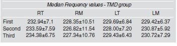
Table 4: Mean values and standard deviation of MF (Hz) of the right temporalis (RT), right masseter (RM), left temporalis (LT) and left masseter (LM) muscles for the first, second and third time intervals examined, in the group with TMD (n=10).
The mean values of MF of the control and TMD groups were compared with muscles (right temporalis (RT), right masseter (RM), left temporalis (LT) and left masseter (LM) and no statistically significant difference was identified in the first three masticatory cycles (p>0.05), in the other three subsequent masticatory cycles (p>0.05) and for the last three masticatory cycles (p>0.05). The Kruskal-Wallis test was applied to compare MF values of the masticatory muscles of control group and TMD group and also did not present statistically significant differences (p>0.05), in all muscles evaluated in any time intervals.
DISCUSSION
Muscle fatigue defined as the inability to maintain an expected force, has been widely investigated in clinical areas. The understanding of the mechanisms involved in the regulation of muscle contraction under conditions of fatigue is of great importance, since fatigue depends on the type of muscle involved, duration of the contraction, level of overload and type of task performed14,15,16. The results of this study show that the values for electromyographic signal amplitude and median frequency do not change significantly during the course of mastication, indicating the absence of muscle fatigue according to the operational definition based on electromyography, in both of the groups studied. One of the factors capable of altering median frequency values is the metabolites accumulation and ions in the interstitial medium, especially protons17. Therefore, although orofacial muscles and the masseter need a greater blood supply, when compared to the biceps and first interosseous muscles18, it is likely that the effect of dynamic activity with successive periods of contraction and relaxation during non-habitual mastication (isotonic contraction), provided adequate blood flow, with the consequent removal of metabolites and ions that cause fatigue. The opposite is observed in isometric contractions, when blood flow is diminished. This finding is associated with the type of muscle fibers that make up the masseter and temporalis muscles. The masseter and temporalis muscles are composed of 58 and 54% type II fibers, respectively, representing a lower oxidative capacity than that of muscles with a predominance of type I fibers19. Upon muscular force used during the phases of jaw closing, although not controlled objectively in this study, the small force that arises in the masticatory activity did not show a relationship with the finding of the present study. Sevensson, Burgaard & Schlosser20 reported that in a group of 11 volunteers without TMD, the contraction sustained with teeth clenching at 10% of the maximum force, maintained for 60 minutes, induced a significant decrease in MF and RMS, besides increasing the sensation of fatigue recorded using a visual analog scale.
When a muscle becomes locally fatigued after repeated contractions, a decrease in the amplitude of the electromyographic signal can be expected. However, in attempt to maintain the level of tension in the muscle, active motor units fire at increasing speed to compensate for the decline in force due to fatigued fibers, resulting in an elevation in amplitude of the electromyographic signal as the muscle fatigues, because of a greater synchrony of firing17. The time necessary to produce fatigue and pain in the mandibular elevator muscles (masseter and temporalis) during teeth clenching was studied by Christensen21 in 14 volunteers with normal occlusion. The mean resistance time of these volunteers was 31 seconds (±11) from the beginning of maximum voluntary contraction till the report of fatigue, which was defined as a subjective event of discomfort.
Although physiological fatigue detected by EMG should precede the subjective reporting of fatigue22 and according to Basmajian and De Luca17, the modifications in the EMG power spectrum during fatigue task should be maximal at the beginning of the task, a period of 15 seconds of masticatory activity was not sufficient to detect signals of fatigue in the recordings obtained. Therefore, the lack of change in MF and RMS values observed in this study can be attributed to two factors: 1) the short period of mastication recording and 2) the dynamic activity which can favor blood flow, reducing the accumulation of protons and metabolites inside the muscle. Based on the results obtained and the experimental conditions of this study, it can be concluded that there are no differences in the electromyographic amplitude (RMS) and median frequency between volunteers with TMD and clinically normal volunteers with regard to a short masticatory activity. Such results can be attributed to dynamic activity which could favor local circulation, and to the individual characteristics of muscle fiber composition and recruitment.
ACKNOWLEDGMENT
The National Council for Scientific and Technological Development (CNPq). Agency linked to the Ministry of Science and Technology (MCT) of Brazil.
1. Okeson JP, de Kanter RJ. Temporomandibular disorders in the medical practice. J Fam Pract. 1996 Oct;43(4):347-356.
2. Inoue-Minakuchi, M. Intramuscular haemodynamic responses to different durations of sustained extension in normal human masseter. Arch Oral Biol 2001;46(7):661-666. [ Links ]
3. Kroon GW, Naeije M. Electromyographic evidence of local muscle fatigue in a subgroup of patients with myogenous craniomandibular disorders. Arch Oral Biol 1992;37(3):215-218. [ Links ]
4. Pinho JC, Caldas, FM, Mora MJ, Santana-Penin U. Electromyographic activity in patients with Temporomandibular disorders. J Oral Rehabil 2000;27:985-990. [ Links ]
5. Cooper BC, Cooper DL. Recognizing Otolaryngology Symptoms in Patients with Temporomandibular Disorders. Cranio 1993;11(04):260-267. [ Links ]
6. De Luca CJ. The use of surface electromyography in biomechanics. J Appl Biomech 1997;13:135-163. [ Links ]
7. Ng JK, Richardson CA, Kippers V, Parnianpour M, Bui BH. Clinical applications of power spectral analysis of electromyographic investigations in muscle function. Man Ther 2001;1(2):99-103. [ Links ]
8. Buzinelli RV, Berzin F. Electromyographic analysis of fatigue in temporalis and masseter muscles during continuous chewing. J Oral Rehabil 2001;28:1-3. [ Links ]
9. Cifrek M, Medved V, Tonković S, Ostojić S. Surface EMG based muscle fatigue evaluation in biomechanics. Clin Biomech (Bristol, Avon). 2009; 327-340.[Epub ahead of print]. [ Links ]
10. Matsumoto T, Ito K, Moritani T. The relationship between anaerobic threshold and eletromyographic fatigue threshold in college women. Eur J Appl Physiol 1991;63:1-3. [ Links ]
11. De Luca CJ. The use of surface electromyography in biomechanics. J Appl Biomech 1997;13(2):135-163. [ Links ]
12. Guevel A, Hogrelj J, Marini J. Fatigue of elbow flexors during repeated flexion – extension cycles: effect of movement strategy. Int J. Sports Med 2000;21:492-498.
13. Dworkin SF, LeResche L. Research diagnostic criteria for temporomandibular disorders: Review, criteria, examinations and specifications, critique. J Craniomandib Disord. 1992;6:301-355. [ Links ]
14. Dimitrova NA, Dimitrov GV. Interpretation of EMG changes with fatigue: facts, pitfalls and fallacies. J Electromyogr Kinesiol 2003;13:13-36. [ Links ]
15. Enoka RM, Stuart DG. Neurobiology of muscle fatigue. J Appl Physiol 1992;72:1631-1648. [ Links ]
16. Latash ML. Chapter 27: Fatigue. In: Neurophysiological basis of movement, Human Kinetics Publishers 2008. [ Links ]
17. Basmajian JV, De Luca CJ. Muscles Alive: Their Functions Revealed by Electromyography. 5th ed. Baltimore, Md: Williams & Wilkins; 1985. [ Links ]
18. Dahlstrom L. Electromyographic studies of craniomandibular disorders: a review of the literature. J Oral Rehabil 1989; 16(01):1-20. [ Links ]
19. Farella M, Van Eijden T, Baccini M, Michelotti A. Task-related electromyographic spectral changes in the human masseter and temporalis muscles. Eur J Oral Sci 2002;110:8-12. [ Links ]
20. Svensson P, Burgaard A, Schlosser S. Fatigue and pain human jaw muscles during a sustained, low-intensity clenching task. Arch Oral Biol. 2001;46:773-777. [ Links ]
21. Christensen LV. Some subjective parameters in experimental toth clenching in man. J Oral Rehabil 1979;6:119-136. [ Links ]
22. Electromyography: Physiology, Engineering and Noninvasive Applications. Chapter: Myoeletric manifestation of muscle fatigue. Merletti R, Parker P, Merletti R, Rainoldi A, Farina D 2004 Wiley-IEEE Press. [ Links ]

