


Serviços Personalizados
Journal

Artigo
 Inglês (pdf)
 Artigo em XML
 Referências do artigo
 Enviar este artigo por email
Indicadores
-
 Citado por SciELO
Links relacionados
-
 Similares em
SciELO
Similares em
SciELO
Compartilhar

 Permalink
Acta Odontológica Latinoamericana
versão On-line ISSN 1852-4834
Acta odontol. latinoam. vol.23 no.1 Buenos Aires abr. 2010
ARTÍCULOS ORIGINALES
Effect of sputter-coating on cracking of root-end surfaces after ultrasonic retrograde preparation - A SEM study of resected root apices and their respective impressions
Clovis M. Bramante1, Ivaldo Gomes de Moraes1, Norberti Bernardineli1, Roberto Brandão Garcia1, Cláudio Urra Pidero2, Ronald Ordinola-Zapata1, Alexandre Silva Bramante1
1Department of Endodontics, Bauru Dental School of Bauru, University of São Paulo, Brazil.
2Private Practice in Concepción, Chile.
CORRESPONDENCE Prof. Dr. Clovis Monteiro Bramante Faculdade de Odontologia de Bauru – USP e-mail: bramante@fob.usp.br
ABSTRACT
The aim of this study was to evaluate in vitro the effect of sputter- coating for SEM analysis on the formation of cracks on root-end surfaces after retrograde cavity preparation with ultrasonic tips. Root-end cavities were prepared with either Satelec S12/90oD diamond-coated or S12/90o non-coated stainless steel retrotips. Impressions were taken before and after retrograde cavity preparation. The resected root apices and their respective impressions were examined using a scanning electron microscope, and the presence, extension and numbers of cracks were recorded after each procedure. The number of cracks observed directly on the ultrasonically prepared root-end surfaces was larger than that observed in their respective impressions taken after root-end cavity preparation, which suggests that cracking was mostly produced by the sputtercoating process required for SEM analysis. Impressions of the root-end cavities prepared with non-coated ultrasonic stainless steel retrotips showed a greater incidence of cracks (3/10 impressions) than those that replicated cavities prepared with diamond-coated retrotips (1/10 impressions). No statistical difference was found between the diamond and stainless steel retrotips.
Key words: Ultrasonic; Root canal therapy; Tooth apex; Microscopy.
RESUMEN
Efecto del metalizado sobre la formación de fracturas en retropreparaciones apicales realizadas con ultrasonido. Estudio con microscopía electrónica de barrido sobre ápices y sus impresiones
El objetivo del presente estudio fue evaluar in vitro el efecto del proceso de metalizacion de la dentina, necesaria para la observacion al microscopio electronico de barrido, sobre la formacion de fracturas dentinarias en retropreparaciones apicales realizadas con ultrasonido. Se realizaron retropreparaciones apicales en 20 piezas dentarias utilizando puntas ultrasonicas diamantadas Satelec S12/90° D (N=10) o puntas no diamantadas Satelec S12/90°. Se tomaron impresiones con silicona antes y despues de que fueran realizadas las retropreparaciones. Los apices radicu - lares y sus respectivas impresiones fueron metalizados y examinadas utilizando un microscopio electronico de barrido. La presencia, extension y numero de fracturas en la dentina fue registrada despues de cada procedimiento. El numero de fracturas observados en la dentina en las retropreparaciones fue mayor que el observado en las impresiones tomadas despues de realizado el procedimiento ultrasonico, lo cual indica que gran cantidad de fracturas en la dentina fueron producidas por el proceso de metalizacion requerido para el analisis al microscopio electronico de barrido y no por el procedimiento ultrasonico. El analisis de las impresiones mostro una mayor incidencia de fracturas cuando se utilizaron las puntas ultrasonicas no diamantadas (3/10) que cuando se utilizaron las puntas ultrasonicas diamantadas (1/10). Sin embargo, la diferencia no fue estadisticamente significativa (p>0.05).
Palabras clave: Ultrasonido; Tratamiento de los conductos radiculares; Cirugia apical; Microscopia.
INTRODUCTION
Endodontic surgery can be defined as the surgical procedure that aims to treat complications unsolved by conventional root canal treatment1,2. Apicoectomy combined with retrograde filling is one of the most widely performed endodontic surgical procedures. The ideal root-end cavity preparation can be described as at least 3-5 mm deep class-I cavity, with walls parallel to the long axis of the root. This regularly shaped cavity should incorporate the root canal anatomy and should retain the retrograde filling material1. The use of ultrasound-activated tips for root-end cavity preparation improves this procedure since less removal of bone tissue is required to gain proper access to the apical region. Also, the resected apical segment can be smaller and less angulated3-5. In addition, ultrasonically prepared root-end cavities can be more conservative than bur-prepared cavities, involving both the canal and the isthmus, allowing better adaptation of the retrofilling material and consequently improving the apical seal. Both non-coated stainless steel and diamond- coated retrotips can be used for root-end cavity preparation6,7,8,10.
Several studies have mentioned the potential of cracking the root-end surface during preparation of retrograde cavities with ultrasound-activated tips12-15. Other studies have not found any evidence of dentin and radicular damage9,16-20. In a clinical study, Sumi et al.11 showed high clinical success rates after the use of ultrasonic root-end preparation for periradicular surgery. Root cracks are classified as complete, incomplete or intradentinal10,14,15,18. A complete crack extends from the root canal to the external root surface; an incomplete crack extends from the root canal to the center of one of the dentinal surfaces; an intradentinal crack is limited to the root dentin, without compromising the canal walls or the external root surface. Root cracks after the use of ultrasound-activated tips have been detected using stereomicroscopy and scanning electron microscopy (SEM), and are the consequence of several reasons, such as the use of dehydrated extracted teeth15,18,21, absence of periodontal ligament13,18,21, power settings of the ultrasound unit5,8,17,21 and sputter- coating of specimens for SEM examination10,18,22. The aim of this study was to evaluate in vitro the effect of sputter-coating on the formation of cracks on the root-end surfaces after retrograde cavity preparation with two ultrasonic tips by SEM examination of the resected root apices and their impressions.
MATERIALS AND METHODS
Twenty fresh extracted human maxillary molars were obtained and stored in 10% formalin buffer solution until use. The palatal root of each tooth was sectioned at the cemento-enamel junction with a water-cooled diamond disk and separated from the crown. Apicoectomy was performed at 3 mm from the apex using a water-cooled low-speed #700 carbide bur positioned perpendicular to the root. The resected apical root segments were replicated with a polyvinylsiloxane impression material (Addflow, SSWhite, Rio de Janeiro, Brazil), obtaining a total of 20 impressions. In half of the teeth (n=10) the root-end cavities were prepared with diamond-coated retrotips S12/90oD (Satelec, Merignac, France). The other teeth were prepared using the S12/90o non-coated stainless steel retrotips (Satelec). Both types of tips were attached to the handpiece of an ultrasonic unit (Jet-Sonic Four Plus; Gnatus, Ribeirao Preto, SP, Brazil) set to the Endo mode with the level of intensity adjusted to 5 (1 to 10 range) under continuous water flow. The depth of the cavities was the same as the cutting end of the ultrasonic retrotips (3 mm). After preparation of the root-end cavities, the resected apical segments were replicated again with the same polyvinylsiloxane impression material (Addflow, SS White, Brazil).
The 20 teeth with the root-end cavities and their respective 40 impressions (20 after apicoectomy and 20 after preparation of the root-end cavities) were mounted on stubs, sputter-coated with gold (Denton Vacuum Inc., Morrestown, NJ, USA) and examined with a scanning electron microscope (JSM, 220A, JEOL Tokyo, Japan) at 15X magnification. The images were analyzed and the presence and number of cracks formed after each procedure were recorded. Cracks extending from the root canal wall that did not include the root surface were considered as incomplete and those that reached the extension of the resected root surface were considered as complete. After the evaluation of the impressions, non-parametric Fisher’s exact test was used to compare the number of roots with the presence of cracks in the coated and non-diamond coated retrotips groups. The level of significance was set at 5%.
RESULTS
Data concerning the presence, extension and number of cracks observed on the specimens ultrasonically prepared and on the impressions taken after apicoectomy and after cavity preparation are shown in Table 1 (diamond-coated ultrasonic tips) and Table 2 (stainless steel ultrasonic tips). In the impressions taken immediately after apicoectomy, only 3 cracks were observed: two in the group prepared with diamond-coated ultrasonic tips and one in the group prepared with stainless steel tips (Tables 1 and 2).
Table 1: Number and extension of cracks on root end surface observed on SEM images of impressions taken after apicectomy and after ultrasonic root-end cavity preparation with diamond-coated ultrasonic tips, and direct SEM images of the root-end surface after cavity preparation and sputter-coating.
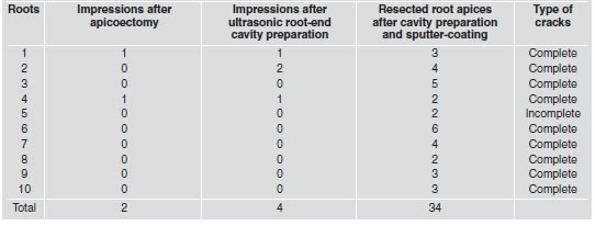
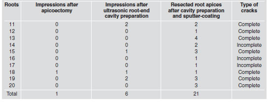
Table 2: Number and extension of cracks on root end surface observed on SEM images of impressions taken after apicectomy and after ultrasonic root-end cavity preparation with non-coated stainless steel ultrasonic tips, and direct SEM images of the root-end surface after cavity preparation and sputter-coating.
Analyses of the impressions gave different results. After root-end cavity preparation with the diamond-coated ultrasonic tips, 4 cracks were observed in the impressions of this group. Considering that two preoperative cracks were noted after apicoectomy in the root-end surface (Table 1), it might be inferred that only 2 new cracks resulted from cavity preparation, both in the same root. In the impressions obtained after use of stainless steel ultrasonic tips, 6 cracks were observed (Table 2). As 1 crack was noted immediately after apicoectomy, 5 cracks resulted from ultrasonic root-end preparation, these cracks were present in 3 roots. No statistical difference was found between the root-end cavity preparation techniques (p>0.05). Direct analyses of the apical surfaces showed that after the sputter-coating process all resected roots presented cracks. There were 34 cracks in the group prepared with the diamond-coated ultrasonic tips and 21 in the group prepared with stainless steel tips. Out of this total, 9 cracks were classified as complete in the group prepared with diamond-coated ultrasonic tips and 7 in the group prepared with stainless steel tips (Tables 1 and 2).
Figure 1 (diamond-coated ultrasonic tips) and Figure 2 (stainless steel ultrasonic tips) show SEM from the impressions of resected root surfaces taken after apicoectomy (A) and after ultrasonic root-end cavity preparation (B), as well as direct images of the respective resected apical root segment (C). Cracking was observed only after sputter-coatig of the specimen.
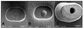
Fig. 1: SEM micrographs of impressions and specimen from the group prepared with diamond-coated ultrasonic tips. A: Impression taken after apicoectomy; B: Impression taken after root-end cavity preparation; C: Resected root apex after rootend cavity preparation and sputter-coating. Root dentin cracking was noted only in the sputter-coated root apex.
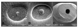
Fig. 2: SEM micrographs of impressions and specimen from the group prepared with stainless steel ultrasonic tips. A: Impression taken after apicoectomy; B: Impression taken after root end cavity preparation; C: Resected root apex after rootend cavity preparation and sputter-coating showing cracks.
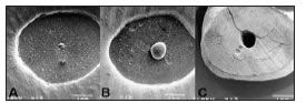
Fig. 3: SEM microphotographs of impressions taken after apicectomy (A) and after retrograde preparation of the rootend cavity (B), as well as an image of the respective resected root-end surface after ultrasonic retrograde preparation with a diamond-coated ultrasonic tip. Cracks were noted after rootend cavity preparation. The number and extension of the root dentin cracks increased after the sputter-coating treatment (C).
DISCUSSION
Previous studies have shown that root-end cavity preparation with conventional burs has a number of difficulties, such as limited access due to radicular anatomy or tooth angulations3-5. Ultrasonic-activated retrotips have been developed to overcome these difficulties and they have been used with high clinical success in retrograde preparation3-5. In spite of the advantages of the ultrasonic retrograde preparation, the potential formation of root dentin microfractures during root-end cavity preparation has been mentioned as a disadvantage of the ultrasonic technique. Saunders et al.12 showed that cracking of the root-end surface was more frequent after ultrasonic root-end preparation than after preparation with a low-speed round bur. This result was also shown by other studies13-15. On the other hand, no cracks could be seen in other studies with ultrasonic root-end preparation methods9,16-20.
The causes of root dentin cracking during in vitro evaluation of ultrasonic retrograde preparation may include: the use of dehydrated extracted teeth, small-sized resected root ends, power settings of the ultrasound unit and sputter-coating process for SEM analysis5,8,10,18,21. As in previous studies, we analyzed SEM images of polyvinylsiloxane impressions of resected root apices after apicoectomy and after retrograde cavity preparation. SEM images of the corresponding root-end surfaces were also studied in order to determine whether root dentin cracks were caused by the ultrasonic preparation or more likely resulted from the specimen processing for SEM analysis10,18,22. The accuracy of the polyvinylsiloxane impressions was confirmed because the root dentin cracks detected in impressions taken after apicoectomy (n=3) were consistently replicated in the impressions taken after the root-end cavity preparation.
An additional advantage of the impression technique is that it enables comparison of the root surfaces before and after root end cavity preparation, and thus discrimination between preoperative and postoperative cracks22. Replicas have been used in other studies of ultrasonic root-end cavity preparation13,17,18,22. In these studies, pouring resin over the impression produced “positive” replicas. “Positive” replicas were not prepared in the present study, because “negative” impressions were considered adequate to observe the occurrence of cracks in the evaluated specimens. The analysis of the sputter-coated resected root-end surfaces showed a great number and extension of cracks in all specimens independently of the type of ultrasound tip used for root-end cavity preparation. These findings demonstrate that most cracks actually resulted from artifacts rather than from ultrasonic root-end cavity preparation. Wucheninch and Meadows5 and Bruyne and De Moor15 analyzed the integrity of root apices of cadaver and extracted teeth after resection and ultrasonic root-end cavity preparation at medium and low ultrasonic power settings at the scaling mode. Extracted teeth showed significantly more root dentin cracks than cadaver teeth, with no differences between the ultrasound intensities. In the present study, medium ultrasonic power setting was used for 2 min, which seemed to minimize the occurrence of microfractures, especially after the use of the diamond-coated ultrasonic tips, as indicated by the analysis of the impressions after root-end preparation, corroborating the findings of Waplington et al.17.
Based on the analysis of the impressions taken before and after root-end cavity preparation, the S12/90D diamond-coated tip produced a lower incidence of cracks (1 out of 10 impressions) than the non-coated stainless steel tip (3 out of 10 impressions). In spite of the fact that all root-end cavities were prepared under water cooling, the results of this study may be attributed to the ability of the diamond-coated tips to abrade the radicular dentin more quickly than noncoated metal tips, thus decreasing heat generation.
CONCLUSIONS
The results of this study showed that cracks observed on dentin surface after ultrasonic root-end pre paration can be artifacts created by the sputtercoating process required for SEM analysis. Impressions of the root-end cavities prior to the SEM analysis are more reliable for evaluating the effect of ultrasonic devices on dentin structure. The non-coated ultrasonic stainless steel retrotips showed a higher incidence of cracks (3/10 impressions) than those cavities prepared with diamondcoated retrotips (1/10 impressions). No statistical difference was found between the two apical preparation techniques.
ACKNOWLEDGMENTS
The authors would like to thank Edimauro de Andrade for his expert technical assistance.
1. Bramante CM, Berbert A. Endodontic surgery. Editora Santos. 2000. [ Links ]
2. Kim S, Kratschman S. Modern endodontic surgery concepts and practice. A review. J Endod 2006;32:601-623. [ Links ]
3. Carr GB. Ultrasonic root-end preparation. Dent Clin North Am 1997;41:541-544. [ Links ]
4. Gutmann JL, Saunders WP, Nguyen l, Guo IY, Saunders M. Ultrasonic root preparation-part 1 SEM analysis. Int Endod J 1994;27:318-324. [ Links ]
5. Wucheninch G, Meadows O, Torabinejad M. Comparison of retrograde cavities prepared by ultrasonic tips and burs (Abstract). J Endod 1993;19:206. [ Links ]
6. Brent PD, Morgan LA, Marshall JG, Baumgartner JC. Evaluation of diamond-coated ultrasonic instruments for root-end preparation. J Endod 1999;25:672-675. [ Links ]
7. Zuolo ML, Perin FR, Ferreira MO, Faria FP. Ultrasonic root-end preparation with smooth and diamond-coated tips. Endod Dent Traumatol 1999;15:265-268. [ Links ]
8. Peters CI, Peters AO, Barbakow F. An in vitro study comparing root-end cavities prepared by diamond-coated and stainless steel ultrasonic retrotips. Int Endod J 2001;34:142-148. [ Links ]
9. Navarre SW, Steiman HR. Root end fracture during rootend preparation: a comparison between zirconium nitride-coated and stainless steel microsurgical ultrasonic instruments. J Endod 2002;28:330-332. [ Links ]
10. Taschieri S, Testori T, Francetti L, DelFarro M. Effects of ultrasonic root-end preparation on resected root surface: SEM evaluation. Oral Surg Oral Med Oral Pathol Oral Radiol Endod 2004;98:611-618. [ Links ]
11. Sumi Y, Hattori JW. Ultrasonic root-end preparation clinical and radiographic evaluation of results. J Oral Maxillofac Surg 1996;54:590-593. [ Links ]
12. Saunders WP, Saunders EM, Gutmann JL. Ultrasonic root-end preparation. Part 2. Microleakage of EBA root-end fillings. Int Endod J 1994;27:325-329. [ Links ]
13. Abedi HR, Van Mierlo BL, Wilder-Smith P, Torabinejad M. Effects of ultrasonic root end cavity preparation on the root apex. Oral Surg Oral Med Oral Pathol Oral Radiol Endod 1995;80:207-213. [ Links ]
14. Layton CA, Marshal JG, Morgan LA, Baumgartner JC. Evaluation of cracks associated with ultrasonic root-end preparation. J Endod 1996;22:157-160. [ Links ]
15. Bruyne MAA, Moor RJG. SEM analysis of the integrity of resected root apices of cadaver and extracted teeth after ultrasonic root end preparation at different intensities. Int Endod J 2005;38:310-319. [ Links ]
16. Lloyd A, Jaunberzins A, Dummer PM, Bryant S. Root-end cavity preparation using the Micromega Sonic retro tip. SEM Analysis. Int Endod J 1996;29:295-301 [ Links ]
17. Waplington M, Lumley PJ, Walmsley AD. Incidence of root surface alteration after ultrasonic retrograde cavity preparation. Oral Surg Oral Med Oral Pathol Oral Radiol Endod 1997;83:387-392. [ Links ]
18. Calzonetti KJ, Iwanowski T, Komorowski R, Friedman S. Ultrasonic root end cavity preparation assessed by an in situ impression technique. Oral Surg. Oral Med Oral Path Oral Radiol Endod 1998;85:210-215. [ Links ]
19. Bernardes RA, Moraes IG, Garcia RB, Bernardineli N, Baldi JV, Victorino FR, Vasconcelos BC, Duarte MAH, Bramante CM. Evaluation of apical cavity preparation with a new type of ultrasonic diamond tip. J Endod 2007;33:484-487. [ Links ]
20. Beling KL, Marshall JG, Morgan LA, Baumgartner JC. Evaluation of cracks associated with ultrasonic instruments for root-end preparation. J Endod 1997;25:323-326. [ Links ]
21. Gray GJ, Steiman HR, Gartner AH. Quality of root-end preparations using ultrasonic and rotary instrumentation in cadavers. J Endod 2000;26:28163 [ Links ]
22. Gondim Jr E, Gomes BP, Ferraz CC, Teixeira FB, Souza-Filho FJ. Effect of sonic and ultrasonic retrograde cavity preparation on the integrity of root apices of freshly extracted human teeth: scanning electron microscopy analysis. J Endod 2002;28:646-650. [ Links ]

