


Servicios Personalizados
Revista

Articulo
 Inglés (pdf)
 Articulo en XML
 Referencias del artículo
 Enviar articulo por email
Indicadores
-
 Citado por SciELO
Links relacionados
-
 Similares en
SciELO
Similares en
SciELO
Compartir

 Permalink
Acta Odontológica Latinoamericana
versión On-line ISSN 1852-4834
Acta odontol. latinoam. vol.24 no.3 Buenos Aires dic. 2011
ARTÍCULOS ORIGINALES
Analysis of hybrid layer thickness, resin tag length and their correlation with microtensile bond strength using a total etch adhesive to intact dentin
Rodolfo B. Anchieta1, Fernanda G. Oliveira2, Renato H. Sundfeld2, Vanessa Rahal2, Lucas S. Machado2, Rodrigo S. Alexandre3, Maria L.M.M. Sundefeld4, Eduardo P. Rocha1
1 Department of Dental Materials and Prosthodontics, São Paulo State University, Faculty of Dentistry of Araçatuba-UNESP, São Paulo, Brazil
2 Department of Restorative Dentistry, São Paulo State University, Faculty of Dentistry of Araçatuba-UNESP, São Paulo, Brazil
3 Department of Restorative Dentistry, University of Guarulhos-UNG
4 Discipline of Biostatistics, São Paulo State University, Faculty of Dentistry of Araçatuba-UNESP, São Paulo, Brazil
CORRESPONDENCE Dr. Sundfeld Renato Rua Jose Bonifacio 1193 Vila Mendonca – Aracatuba / SP, Brasil, 16015-050 e-mail: sundfeld@foa.unesp.br
ABSTRACT
The objective of this study was to evaluate the use of a two-step total etch and rinse adhesive, the correlation between the hybrid layer thickness (HL) and bond strength (BS), and between resin tag length (RT) and bond strength in the same teeth, and also to evaluate the fracture patterns of the tested specimens. Ten human molars were used for the restorative procedure and then sectioned in two halves (mesio-distally). The materials used were Adper™ Single Bond 2, 3M ESPE, Ultra etch gel, Ultradent and Filtek™ Z250, 3M ESPE. One half were utilized to measure the HL thickness and RT length through light microscopy analysis (400x), and the other half was subject to a microtensile test to measure the BS. The fractured surfaces were analyzed by scanning electron microscopy and fracture patterns classified. The Pearson correlation test was applied (p=0.05). The results of the analyses of each specimen then were correlated: mean HL thickness = 4.39(0.48) μm, mean length of RT = 9.94(1.69) μm, mean BS = 23.98(10.24) MPa. A statistically significant correlation between HL thickness and bond strength was found (r=0.93). The two step etch and rinse adhesive system, showed a strong correlation between HL thickness and bond strength. The most common fractures were adhesive, followed by cohesive in resin.
Key words: Dentin bonding agents; Dentin; Dental adhesives.
RESUMO
Análise da espessura da camada híbrida, comprimento dos tags e sua correlação com a resistência de união a microtração, utilizando um sistema adesivo de condicionamento ácido total em dentina hígida
Objetivos: O objetivo deste estudo foi avaliar utilizando um sistema adesivo de 2 passos de condicionamento ácido total, a correlação entre a espessura da camada híbrida (CH) e a resistência de união (RU), e entre o comprimento dos tags resinosos (TR) e a RU em um mesmo dente utilizando, assim como o padrão de fratura dos espécimes testados. Material e método: Dez molares humanos foram utilizados para o procedimento restaurador e seccionados em duas metades (mésio-distal). Os materiais utilizados foram Adper ™ Single Bond 2, 3M ESPE, gel Ultra ™ etch, Ultradent e Filtek Z250, 3M ESPE.Uma metade foi utilizada para a microscopia de luz (400x) para se obter a espessura da CH e o comprimento dos TR, e a outra metade foi submetida ao teste de microtração para obtenção da RU. As superfícies fraturadas foram analisadas por microscopia eletrônica de varredura e os padrões de fratura classificados. O teste de correlação de Pearson foi aplicado p=0.05. Resultados: Os resultados das análises de cada espécime (CH, TR e RU), em seguida, foram correlacionados: CH espessura média = 4,39(0,48) μm, comprimento médio dos TR = 9,94(1,69) μm, média da RU= 23,98(10,24) MPa. Correlação estatisticamente significativa foi encontrada entre a espessura da CH e RU (r=0,93). Conclusão: o sistema adesivo de condicionamento ácido total de dois passos mostrou forte correlação entre a espessura da camada híbrida e resistência de união. Os tipos de fraturas mais comuns foram a adesiva, seguida da coesiva na resina.
Palavras Chave: Agentes de união dental; Dentina; Adesivos dentais.
INTRODUCTION
The bonding capacity of restorative materials to dentin has been enhanced by the evolution of adhesive systems in the past decade1. The efficiency of adhesive systems is caused by the formation of a dentin–adhesive interface (d/a interface) with sufficient physical and mechanical properties2 to allow the restorations to withstand the masticatory forces and conditions inherent to the oral cavity3. The efficiency of bonding to dentin mostly depends on micromechanical retention promoted by resin infiltration in partially demineralized dentin, with consequent hybrid layer (HL)4,5 and resin tag (RT) formation inside the dentin tubules6-8 responsible for producing high bond strength. However, some authors have found variations in the thickness of the HL and the length of RT, as well as different morphologies of the resultant adhesive interface depending on the type of adhesive system used and variation on bond strength values in laboratory studies9.
The introduction of the microtensile test10 allowed the true bond strength of adhesives systems, to be measured by using, cross-sectional areas of each specimen. Specimens with a small cross-sectional area allows better stress distribution, and better specimen homogeneity, reducing the occurrence of defects, consequently reducing the occurrence of cohesive fractures in the substrate, and finally contributing to the measurement of actual bond strength.6 Furthermore, they make it possible to evaluate the bond strength (BS) in different regions on the same substrate and investigate bond strength on irregular surfaces11. Even though significant dentin hybridization is necessary for the achievement of high bond strength values, few studies have addressed and correlated RT length and HL thickness with bond strength values to dentin in the same teeth12-14.
The objective of this study was to evaluate the correlation between hybrid layer thickness and bond strength and betweem resin tag length and bond strength in the same teeth, using a 2- step etch and rinse adhesive system to intact dentin. In addition, the fractured surfaces were analyzed by scanning electron microscopy and the fracture patterns were classified. The null hypothesis was there is no correlations between the hybrid layer thickness and bond strength or between resin tag length and bond strength.
MATERIALS AND METHODS
Selection of teeth and study design
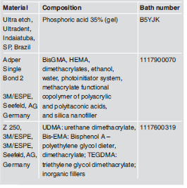
The study was conducted on 10 intact human molars, which were stored in distilled water until use, (up to 6 months after extraction). The study was revised and approved by the Institutional Review Board. The study used the twostep total-etch adhesive system Adper™ Single Bond 2 (3M ESPE, 3M/ESPE, Seefeld, AG, Germany), as well as Ultra etch gel (Ultradent, Indaiatuba, SP, Brazil), composed of 35% phosphoric acid, and composite resin (Filtek™ Z250, 3M ESPE, 3M/ESPE, Seefeld, AG, Germany) (Table 1).
Table 1: Materials employed in this study (compon e nts, manufacturers).
Preparation of teeth
The occlusal enamel was removed with a diamond disc (IsoMetR Diamond Wafering Blade, Buehler Ltd, Lake Bluff, IL, USA), three millimeters up to dentin-cement junction under constant irrigation connected to a hard-tissue microtome (IsoMetR 2000, Buehler Ltd, Lake Bluff, IL, USA), followed by wearing down the surface on a polishing machine (Fortel Ltda, Sao Paulo, SP, Brazil) with silicon-carbide sandpaper grit 320 (Buehler Ltd, Lake Bluff, IL, USA) under irrigation, which exposed the dentin. Thereafter, a standardized smear layer was created with silicon-carbide sandpaper grit 600 (Buehler Ltd, Lake Bluff, IL, USA), also on the polishing machine, under continuous irrigation for 30 seconds (Fig. 1).

Fig. 1: Diagram showing the sequence of preparation of specimens for light microscopy analysis, microtensile test, and scanning electron microscopy analysis. A – Molar teeth; B – Sectioning of tooth crown to expose dentin tissue; C – Exposed dentin tissue; D – Bonding procedures; E – Sectioning of crown into two halves; F – One half was embedded in paraffin to prepare histological sections and stained by the Brown and Brenn method for analysis under a light microscope; G – Sectioning into a sticks with mean cross-sectional area of 1 mm2; H – Microtensile testing; I – Fractured sticks and analysis of fracture patterns under scanning electron microscope.
Bonding Procedures
The specimens were submitted to acid etching of dentin with 35% phosphoric acid gel (Ultra etch, Ultradent, Idaiatuba, SP, Brazil) applied for 15 seconds on the dentin. Then the cavities were carefully rinsed with water for 15 seconds and dried. After drying, the dentin surface should have a moist appearance. To achieve this clinical effect, the dentin tissue was protected with a small cotton pellet while the etched cavities were dried. The two-step adhesive (Adper Single Bond 2, 3M ESPE, 3M/ESPE, Seefeld, AG, Germany) was applied with a brush (MicrobrushR Intl, Grafton, WI, USA) on the dentin surface, which was allowed to rest for 30 second for the solvents to evaporate as recommended by the manufacturer, and, then, light-cured for 20 seconds with a light-curing unit (Ultralux Lens, Dabi Atlante, Ribeirao Preto, SP, Brazil) at an intensity of 450 mW/cm2. A Filtek Z250 (3M ESPE, 3M/ESPE, Seefeld, AG, Germany) composite resin block (shade A2) measuring nearly 4 mm was applied incrementally in a total of approximately 10 increments to the surface in 4 layers, which were light-cured individually for 40 seconds. Bonding was performed under controlled environmental conditions of 22°C with 45% to 55% humidity. Each prepared tooth was sectioned mesiodistally with a diamond disc under constant irrigation on a sectioning machine (IsoMet 2000, Buehler, Lake Bluff, IL, USA) to create two halves (buccal and lingual). Teeth halves were randomly selected by a blinded operator for optical microscopy analyses (HL thickness and RT length) or microtensile bond strength.
Light Microscopy Analysis
The tooth halves used for optical microscopy were decalcified in a solution containing 50% formic acid and 20% sodium citrate, which was changed every 5 days; complete decalcification of each specimen was checked radiographically15, and it was achieved after 3 months. This process completely removed the enamel, leaving only the demineralized dentin tissue, which was the subject of this study. After decalcification, the restorations were removed carefully and then embedded in paraffin14.
Then, the specimens were sectioned longitudinally through their crowns into slices 6 μm thick and mounted on glass slides. Fifteen slides of each specimen containing approximately six sections each were se-lected by systematic sampling, with an interval proportional to the number of sections achieved for each specimen16. These sections were stained with Brown and Brenn stain, after which the better histological section of each, which showed the best stained hybrid layer and tags, was analyzed on under a light microscope (Axiophot, Zeiss DSM-940 A, Carl Zeiss MicroImaging Inc, Thornwood, NY, USA) at 400x magnification, with a 40/075 micrometric eyepiece (Fig. 2). The HL and RT of each section were measured carefully, ensuring that the entire extension of the histological section was analyzed by a single, calibrated examiner. Three measurements were taken of each section, for each factor analyzed. Consequently, for each slide selected, the thickness of the hybrid layer and the length of the resin tags corresponded to the mean of the three measurements performed. Thus, 15 mean values, of the three measurements performed, were obtained for each specimen, for both the HL and the RT14.
Fig. 2: Light microscope image (400X magnification), revealing hybrid layer and resin tags formation. HL = hybrid layer; RT = tags.
Microtensile Test
The other halves were submitted to the microtensile bond strength test14. The root ends of these specimens were attached on an acrylic block connected to the hard-tissue sectioning machine (IsoMet 2000, Buehler, Lake Bluff, IL, USA), to allow sectioning transversely and longitudinally to the dentin–composite adhesive interface. They were sectioned with a diamond disc under water cooling, creating stick-shaped specimens with an approximate transverse section area of tooth– resin interface of 1 mm2. All specimens were prepared after storage in distilled water at 37°C for 24 hours.
Then, the specimens were attached individually by their ends to a microtensile device on the testing machine (Vitrodyne V1000, Chatillon Bros, Greensboro, NC, USA), with aid of cyanoacrylate glue (Dentisply, Sankin KK, Japan). The tensile load was applied perpendicularly to the adhesive interface at a crosshead speed of 0.5 mm/min until rupture of the specimens. After rupture, the specimens were removed from the device with aid of a blade. The transverse section surface area on each fracture plane was measured with a digital pachymeter (Digimess, Shinko Precision Gaging, LTD, China). The dentin side of failed specimens was sputter coated with gold (Balzers SCD 050, Balzers Union, Balzers, Liechtenstein) and observed under SEM (JSM 5600 LV, Jeol Inc., Peabody, MA, USA). Photomicrographs of a representative area of the surface were taken at 100X and 1000X magnification. The fracture patterns were classified as adhesive, cohesive in dentin, cohesive in composite, or mixed if more than one structure was involved in the fracture. The bond strength of the specimens was calculated as the ratio between the load recorded at fracture (kgf) and the measurement of the cross-sectional area (cm2), expressed in MPa. These values revealed the bond strength of each specimen.
Statistical analysis of data
The values obtained for each factor analyzed (HL, RT, and bond strength), corresponding to each specimen were analyzed statistically by Pearson’s correlation test, between the bond strength values and measurements of HL thickness and RT length. The test was applied at a significance level of p=0.05.
RESULTS
After the laboratory procedures, the mean values of hybrid layer thickness was 4.39 (0.48) μm. The mean resin tag length was 9.94 (1.69) μm. Under microtensile tests the means values of bond strength was 23.98 (10.24) MPa. All the specimens fractured during the microtensile test. Table 2 shows the results for these variables.
Table 2: Values for hybrid layer thickness (μm), resin tag length (μm) and bond strength (MPa).

The fractured specimens showed 41% adhesive fracture (Fig. 3), followed of cohesive fracture in resin (34%). Mixed fractures were 25%. No dentin fracture was observed. A statistically significant correlation between the HL and microtensile bond strength (r=0.93) was found (Fig. 4) at 5% level. However, no correlation was found between RT and microtensile bond strength (r=0.007).

Fig. 3: Scanning electron microscopy analysis of adhesive fracture. A. Lower magnification (x100). B. Higher magnification (x1,000) of fracture area on the base of hybrid layer. C. Higher magnification (x1,000) showing a adhesive fracture pattern.

Fig. 4: Correlation between hybrid layer thickness and microtensile bond strength of Adper Single Bond 2. As R2=0.86, the trend line indicates the linear regression equation which allows the estimateion of a value of one variable when the other variable is known.
DISCUSSION
This study evaluated the ability of Adper Single Bond 2 to infiltrate dentin tissue and create a HL, RT as well as its bond strength to intact dentin tissue. Light microscopy analysis allowed the assessment and measurement of the thickness of the HL and length of RT in an extensive dentin area within the same tooth, achieving results consistent with other reports15,16. The HL thickness values were similar to previous studies that confirmed the high degree of hybridization of the adhesive system compared to other total etch adhesivesl3,17,18 and some self-etching adhesive systems3,18,19. The usable thickness of the hybrid layer was probably caused by the complete removal of the smear layer and smear plug followed by dentin demineralization by phosphoric acid etchant, which exposed a large number of collagen fibers4,20. The pathways for the infiltration of resin material into the etched intertubular dentin, which correspond to the peripheral spaces around the collagen fibers, favor the formation of a thick hybrid layer and impair differentiation between HL and RT within it15,16.
With regard to the RTs, the infiltration of adhesive into the dentinal tubules for formation of the tags (Table 2) was not as significant as observed for other adhesive systems8,21. This could be because the higher viscosity of the adhesive system used might have reduced infiltration of the resin material into the dentinal tubules22. The concentration of fillers in the openings of tubules may reduce the infiltration into the tubules22. Moreover, several authors have demonstrated that a specific polyalkenoic copolymer does not dissolve in the adhesive’s solution, leading to the production of many globules within the polymer of the adhesive layer23,24. However, the main benefit of this filled adhesive could be to act as a stress-absorbing layer within the HL, in addition to the better mechanical properties of the hybrid layer24-26. This may have been the reason for the strong correlation between HL thickness and bond strength achieved. Furthermore, the correlation was achieved due high-quality HL formed that strongly bonding the resin material to tooth substrate27.
However, in another study, no correlation between the HL and bond strength was found using a self-etch adhesive system (Adper Prompt L-Pop) 14, although the created HL of this agent were thick, as found in the present study for the two- step etch and rinse adhesive. According to the authors, this may have occurred due the poor quality of the created HL14. Using finite element analysis, others authors showed that for self-etch adhesives, a thick HL contributes to high stress concentration in dentin/adhesive interface28. Additionally, some self-etch adhesives create poor quality HL and are prone to imperfections in HL, such as voids, leading to the concentration of high stress levels inside the HL, suggesting that this these sites are criticals to crack initiation and propagation29, 30. The present study revealed the influence of the hybrid layer on the bond strength for the two- step total etch adhesive system Adper Single Bond 2. Regression analysis revealed a strong correlation between thickness of HL and bond strength, allowing the assumption that a thicker HL provides higher bond strength for this adhesive system.
However, the present study did not reveal correlation between the length of RT and bond strength. The null hypothesis can therefore be partially rejected, since the hybrid layer thickness and bond strength showed a strong correlation. This lack of interaction may be related to application of the microtensile test on the long axis of resin tags, which may have reduced the mechanical interaction of RT29. Moreover, formation of an intratubular hybrid layer was extremely small or even absent, due the highly mineralized peritubular dentin27. In most instances, it was present only on the tubule opening, without great influence on bond strength29.
Another explanation may be related to the resin composition of the tags. When a simplified adhesive, such as Adper Single Bond 2, is applied on etched wet environment dentin tissue, there is deeper migration of molecules with lower molecular weight, ie, the hydrophilic monomers32. According to these authors, the resin tags follow the same pattern (i.e., most resin tags are formed by monomers of low molecular weight), which cure weakly, further reducing their contribution on bond strength32. This was demonstrated by the analysis of the fracture patterns for this adhesive; most adhesive fractures occurred on the base of the HL or within the HL and few cohesive fractures in dentin were observed, demonstrating the lack of influence of RT on bond strength. Within the limitations of the study it ispossible to conclude that the two- step total etch adhesive system used exhibited positive correlation between HL thickness and microtensile bond strength; however did not show a correlation between resin tags and microtensile bond strength. The majority of fractures were adhesive, followed by cohesive in resin.
ACKNOWLEDGEMENTS
The authors would like to thank FAPESP (#06/52520-4) for the financial support.
1. Yeşilyurt C, Bulucu B. Bond strength of total-etch dentin adhesive systems on peripheral and central dentinal tissue: a microtensile bond strength test. Contemp Dent Pract 2006;7:26-36. [ Links ]
2. Wang Y, Spencer P. Hybridization efficiency of the adhesive/ dentin interface with wet bonding. J Dent Res 2003; 82:141-145. [ Links ]
3. Koshiro K, Sidhu SK, Inoue S, Ikeda T, Sano H. New concept of resin-dentin interfacial adhesion: the nano interaction zone. J Biomed Mater Res B Appl Biomater 2006;77:401-408. [ Links ]
4. Van Meerbeek B, De Munck J, Yoshida Y, Inoue S, Vargas M, Vijay P, Van Landuyt K, Lambrechts P, Vanherle G. Buonocore memorial lecture. Adhesion to enamel and dentin: current status and future challenges. Oper Dent 2003;28:215-235. [ Links ]
5. Van Landuyt KL, Snauwaert J, De Munck J, Peumans M, Yoshida Y, Poitevin A, Coutinho E, Suzuki K, Lambrechts P, Van Meerbeek B. Systematic review of the chemical composition of contemporary dental adhesives. Biomaterials 2007;28:3757-3785. [ Links ]
6. Pashley DH, Sano H, Ciucchi B, Yoshiyama M, Carvalho RM. Adhesion testing of dentin bonding agents: a review. Dent Mater 1995;11:117-125. [ Links ]
7. van Meerbeek B, Vargas M, Inoue S, Yoshida Y, Peumans M, Labrechets P, VanHerle G. Adhesives and cements to promote preservation dentistry. Oper Dent 2001;26: 119-144. [ Links ]
8. Gregoire G, Millas A. Microscopic evaluation of dentin interface obtained with 10 contemporary self-etching systems: correlation with their pH. Oper Dent 2005;30: 481-491. [ Links ]
9. Vargas MA, Cobb DS, Denehy GE. Interfacial micromorphology and shear bond strength of single-bottle primer/ adhesives. Dent Mater 1997;13:316-324. [ Links ]
10. Sano H, Shono T, Sonoda H, Takatsu T, Ciucchi B, Carvalho R, Pashley DH. Relationship between surface area for adhesion and tensile bond strength-evaluation of a microtensile bond test. Dent Mater 1994;10:236-240. [ Links ]
11. Pashley DH, Carvalho RM, Sano H, Nakajima M, Yoshiyama M, Shono Y, Fernandes CA, Tay F. The microtensile bond test: a review. J Adhes Dent 1999;1:299-309. [ Links ]
12. Pioch T, Stotz S, Buff E, Duschner H, Staehle HJ.. Influence of different etching times on hybrid layer formation and tensile bond strength. Am J Dent 1998;11:202-206. [ Links ]
13. Prati C, Chersoni S, Mongiorgi R, Pashley DH. Resin infiltrated dentin layer formation of new bonding systems. Oper Dent 1998;23:185-194. [ Links ]
14. Oliveira FG, Anchieta RB, Rahal V, Alexandre RS, Machado LS, Sundefeld MLMM, Giannini M, Sundfeld RH. Correlation of hybrid layer and resin tags with the microtensile bond strength of a self-etch adhesive system. Acta Odontol Latinoam. 2009;22:177-181. [ Links ]
15. Sundfeld RH, Valentino TA, de Alexandre RS, Briso AL, Sundefeld ML. Hybrid layer thickness and resin tag length of a self-etching adhesive bonded to sound dentin. J Dent 2005;33:675-681. [ Links ]
16. Sundfeld RH, da Silva AM, Croll TP, de Oliveira CH, Briso AL, de Alexandre RS, Sundefeld ML. The effect of temperature on self-etching adhesive penetration. Compend Contin Educ Dent 2006;27:552-557. [ Links ]
17. Machado LS, Sundfeld RH, Cardoso JD, Oliveira FG, da Silva AP, Delicio G, de Alexandre RS, Sundefeld MLMM. Observation of tags and hybrid layer of a single bottle conventional adhesive system and a self-etching adhesive system, on sound dentin. Acta Odontol Latinoam 2009;22: 183-189. [ Links ]
18. Arrais CAG, Giannini M. Morphology and thickness of the diffusion of resin through demineralized or unconditioned dentinal matrix. Pesqui Odontol Bras 2002;16:115-120. [ Links ]
19. Waidyasekera PG, Nikaido T, Weerasinghe DD, Tagami Jl. Bonding of acid-etch and self-etch adhesives to human fluorosed dentine. J Dent 2007;35:915-922. [ Links ]
20. Reis A, Grandi V, Carlotto L, Bortoli G, Patzlaff R, Rodrigues Accorinte Mde L, Dourado Loguercio A. Effect of smear layer thickness and acidity of self-etching solutions on early and long-term bond strength to dentin. J Dent 2005; 33:549-559. [ Links ]
21. Gregoire GL, Akon BA, Millas A. Interfacial micromorphological differences in hybrid layer formation between water- and solvent based dentin bonding systems. J Prosthet Dent 2002;87:633-641. [ Links ]
22. Can Say E, Nakajima M, Senawongse P, Soyman M, Ozer F, Ogata M, Tagami J. Microtensile bond strength of a filled vs unfilled adhesive to dentin using self-etch and total-etch technique. J Dent 2006;34:283-291. [ Links ]
23. Van Meerbeek B, Conn LJ Jr, Duke ES, Eick JD, Robinson SJ, Guerrero D. Correlative transmission electron microscopy examination of nondemineralized and demineralized resin– dentin interfaces formed by two dentin adhesive systems. J Dent Res 1996;75:879-888.
24. Van Meerbeek B, Willems G, Celis JP, Roos JR, Braem M, Lambrechts P, Vanherle G. Assessment by nano-indentation of the hardness and elasticity of the resin-dentin bonding area. J Dent Res 1993;72:1434-1442. [ Links ]
25. Unterbrink GL, Liebenberg WH. Flowable resin composites as “filled adhesives”: literature review and clinical recommendations. Quintessence Int 1999;30:249-257.
26. Labella R, Lambrechts P, Van Meerbeek B, Vanherle G. Polymerization shrinkage and elasticity of flowable composites and filled adhesives. Dent Mater 1999;15:128-137. [ Links ]
27. Nakabayashi N, Pashley DH. Hybridization of Dental Hard Tissue. Tokyo: Quintessence Publishing Co; 1998:8-9. [ Links ]
28. Anchieta RB, Rocha EP, Ko CC, Sundfeld RH, Martin Junior M, Archangelo CM. Localized mechanics of dentin self-etching adhesive system. J Appl Oral Sci 2007;15:321-6. [ Links ]
29. Martini A, Anchieta RB, Rocha EP, Freitas Junior AC, Almeida EO, Sundfeld RH, Luersen MA. Influence of voids in the hybrid layer based on self-etching adhesive systems. A 3-D FE analysis. J Appl Oral Sci 2009;17:19-26. [ Links ]
30. Anchieta RB, Rocha EP, Sundfeld RH, Martin Junior M, Giannini M, Reis FA. Micromechanics of dentin/adhesive interface in function of dentin depth: 3D finite element analysis. Int J Clin Dent 2011;4:199-210. [ Links ]
31. Misra A, Spencer P, Marangos O, Wang Y, Katz JL. Micromechanical analysis of dentin/adhesive interface by the finite element method. J Biomed Mater Res B Appl Biomater 2004;70:56-65. [ Links ]
32. Spencer P, Wang Y. Adhesive phase separation at the dentin interface under wet bonding conditions. J Biomed Mater Res 2002;62:447-456. [ Links ]

