


Services on Demand
Journal

Article
 English (pdf)
 Article in xml format
 Article references
 Send this article by e-mail
Indicators
-
 Cited by SciELO
Related links
-
 Similars in
SciELO
Similars in
SciELO
Share

 Permalink
Acta Odontológica Latinoamericana
On-line version ISSN 1852-4834
Acta odontol. latinoam. vol.25 no.2 Buenos Aires Oct. 2012
ARTÍCULOS ORIGINALES
Correlation between hybrid layer thickness, resin tag length and microtensile bond strength of a self-etching adhesive system
Vanessa Rahal1, Fernanda G. de Oliveira1, André L.F. Briso1, Paulo H. dos Santos2, Maria L.M.M. Sundefeld3, Renato H. Sundfeld1
1 Department of Restorative Dentistry, Araçatuba Dental School, São Paulo State University, Araçatuba, São Paulo, Brazil.
2 Department of Dental Materials and Prosthodontics, Araçatuba Dental School, São Paulo State University, Araçatuba, São Paulo, Brazil.
3 Department of Community and Preventive Dentistry, Araçatuba Dental School, São Paulo State University, Araçatuba, São Paulo, Brazil.
CORRESPONDENCE Dr.Vanessa Rahal Discipline of Restorative Dentistry Sao Paulo State University. Rua Jose Bonifacio 1193, Sao Paulo Sao Paulo, Brazil.16015-050 Email: vanessarahal@hotmail.com
ABSTRACT
The purpose of this study was to evaluate the correlation between the hybrid layer thickness, resin tag length and resin bond strength of a self-etching adhesive system to sound dentin tissue "in vivo". After performing restorative procedures and tooth extractions, ten specimens were sectioned in a mesiodistal direction. One dental section was used for light microscope analysis, in which both the resin tag length and hybrid layer thickness were measured, while the other section was analyzed using a microtensile test (0.5 mm/min). The fractured surface of the latter section was characterized using a stereoscopic magnifying glass (40× magnification). The results were subject to statistical analysis using the Pearson Correlation Test (α = 0.05). The hybrid layer thickness, resin tag length and resin bond strength mean values were 2.19 μm (0.34), 4.34 μm (0.28) and 9.73 MPa (5,55), respectively. In addition, correlation tests between the resin tag length and the resin bond strength (r=0.014) and also between the hybrid layer thickness and bond strength (r=0.43), showed no statistically significant correlation. The microtensile bond strength of Adper Prompt L Pop self-etching adhesive system does not depend on hybrid layer thickness or resin tag length.
Keywords: Dentin bonding agents; Tensile Strength; Polarized Light Microscopy.
RESUMO
Correlação entre a espessura da camada híbrida, comprimento dos prolongamentos resinosos e resistência adesiva à microtração de um adesivo autocondicionante
A proposta deste estudo foi avaliar a correlacao entre a espessura da camada hibrida e comprimento dos tags com a resistencia adesiva de um sistema adesivo auto-condicionante, quando aplicado em dentina higida. Depois da realizacao dos procedimentos restauradores, dez especimes foram seccionados ao meio na direcao mesio-distal. Uma metade foi utilizada para analise em microscopia optica, onde os comprimentos dos tags e espessura da camada hibrida foram mensuradas, a outra metade foi analisada por meio do teste de microtracao (0,5 mm/min). As superficies de fratura foram classificadas com o uso de lupa estereoscopica (aumento de 40×). Os resultados foram submetidos a analise estatistica por meio do Teste de Correlacao de Pearson (α = 0,05). Os valores da espessura da camada hibrida, comprimento dos tags resinosos e resistencia adesiva foram obtidas, sendo 2,19 μm (0,34), 4,34 μm (0,28) e 9,73 MPa (5,55), respectivamente. Alem disso, o teste de correlacao entre os comprimentos dos tags resinosos e a resistencia adesiva (r=0,014) e a espessura da camada hibrida e a resistencia adesiva (r=0,43), nao mostraram correlacao estatistica. O teste de microtracao do adesivo auto-condicionante Adper Prompt L Pop nao apresentou correlacao com a espessura da camada hibrida e o comprimento dos tags resinosos.
Palavras-chave: Adesivos dentinarios; Resistencia a tracao; Microscopia de luz polarizada.
INTRODUCTION
Progress in restorative techniques, particularly esthetics, has led manufacturers and researchers to seek improvement in the adhesive and resinous composite systems. The advent of acid etching has provided excellent adhesive bonding of resinous materials to dental enamel1. Dentin, in contrast, is a more complex substrate, and bonding to it continues to pose a challenge, despite recent progress in adhesive techniques2.
Nakabayashi3 and other authors observed that the process of adhesive bonding to acid-etched dentin is mainly of micromechanical nature, and concluded that it is greatly improved by removing the smear layer from the dentin4,5. More recently, new materials and techniques for acid etching dental tissues have been created, for example, the self-etching adhesive systems6,7, which can etch both enamel and dentin, and simultaneously infiltrate their adhesive monomers 8 into the micropores of the enamel and the demineralized collagen matrix. Among the self-etching adhesives, there is the onestep type, considered more aggressive and capable of forming a hybrid layer similar in thickness to those formed with conventional adhesives9,10. Laboratory studies with self-etching adhesive systems have shown the importance of the resin tags and hybrid layer for obtaining an adhesive bond to dentinal tissue7,11. Among the ways of analyzing these resinous structures, some papers report that optical microscope analysis provides consistent information on large areas of the bond interface12,13. On the other hand, Sano et al., in 199414, described the possibility of observing the behavior of adhesive systems by using the microtensile test, claiming that among other advantages, it allows the distribution of forces in a more homogeneous manner throughout the entire sample surface; thereby reducing the incidence of cohesive fractures and making it possible to obtain real bond strength values.
Studies on the bond strength of self-etching adhesive systems by application of the microtensile test have been performed15,16, but few have correlated the values obtained with the length of the resin tags and hybrid layer thickness. Therefore, the aim of this study was to analyze the correlation of the length of resin tags and the thickness of the hybrid layer with the microtensile bond strength of a selfetching adhesive system, when applied to dentinal tissue "in vivo". The null hypothesis tested was that the bond strength was not influenced by the thickness of the hybrid layer and length of resin tags.
MATERIALS AND METHODS
Specimen Preparation and Restorative Procedures
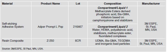
After the project had been submitted to and approved by the proper Research Ethics Committee, ten third molars indicated for extraction were selected from students of the dental undergraduate course. After dental prophylaxis, local anesthesia of the operating field was performed, in order to make wide Class I type cavity preparations involving practically the entire occlusal face, to a depth of 4 mm, standardized by the length of the active part of the 1094 diamond bur (KG Sorensen Industria e Comercio Ltda., Barueri, Sao Paulo, Brazil) used. The diamond bur was mounted in a high-speed handpiece, and used under water and air cooling (MS 350 - Dabi-Atlante, Ribeirao Preto, Sao Paulo, Brazil). A self-etching, one-step adhesive system Adper Prompt L Pop (3M/ESPE, St Paul, MN, USA) and resin composite Z-250 (3M/ESPE, St Paul, MN, USA) were used. Their compositions are provided in Table 1.
Table 1: Materials used in the study.
The ten teeth were divided into three groups according to the type of analysis to be performed on dentinal tissue, considering the following factors: resin tags, hybrid layer and microtensile bond strength (Table 2).
Table 2: Study Groups.

After the cavities had been washed with water and dried with smooth air jets, the adhesive system Adper Prompt L Pop (3M ESPE Dental Products, St Paul, MN, USA) was applied according to the manufacturer's instructions, i.e., under pressure on the dentinal surface for 15 seconds and dried with smooth air jets for 5 seconds. Then, after the applicator had been wet again, a second layer of adhesive was applied and following another application of air jets, it was light-activated for 10 seconds with an Ultralux halogen light source (Dabi Atlante, Ribeirao Preto, Sao Paulo, Brazil) at an intensity of 450 mW/cm2. At this time, the cavities received the application of resin composite Z-250 (shade A2) (3M ESPE Dental Products, St Paul, MN, USA) in layers, starting by inserting the resinous material against the vestibular and pulp walls, and then against the lingual and pulp walls until the entire cavity was filled. Each layer was light-activated for 40 seconds. All the restorations were finished with diamond burs of the Gold Series No.1190F (K.G. Sorensen Industria e Comercio Ltda., Barueri, Sao Paulo, Brazil), followed by the use of Enhance siliconized burs (Dentsply - Industria e Comercio Ltda., Petropolis, Rio de Janeiro, Brazil).
Immediately after this, the restored teeth were carefully extracted, taking every precaution necessary not to compromise the restored area. Finally, the teeth were cleaned and sectioned in the mesio-distal direction, under constant irrigation, in a metallographic cutter ISOMET 1000 (Buehler, Lake Bluff, IL, EUA), to obtain two sections - vestibular and lingual.
Optical Microscopy Analysis
The dental sections used for optical microscopy were decalcified in a solution containing equal proportions of 50% formic acid and 20% sodium citrate. Complete decalcification of the specimen was verified by taking radiographs13,17,18. After decalcification, the restorations were carefully removed, so that the remaining organic material could be embedded in paraffin. The specimens were sectioned in the longitudinal direction of the clinical crown to a thickness of six micrometers, and sequentially mounted on glass slides. Fifteen slides of each specimen were selected, maintaining an interval proportional to the number of cuts obtained of each specimen. After staining the slides by the Brown & Brenn method, the best histological sections of each slide were analyzed by Axiophot optical microscopy (ZEISS DSM-940 A, Oberkochen, Baden-Wurttemberg, Germany) at 1000× magnification, with a 100×/1.30 micrometric eyepiece (Fig.1). The measurements of the hybrid layer and resin tags were taken after meticulous analysis of the entire extent of the histological section with the aid of the software AxioVision Software Real (Version 4.3; Carl Zeiss MicroImaging Inc., Thornwood, NY, USA). For each factor under analysis, three measurements were obtained in each section, in which the resin tags analyzed and measured belonged to the same region of the hybrid layer analyzed and measured. Consequently, for each selected slide, the thickness of the hybrid layer and length of the resin tags would correspond to the mean of the three measurements taken. Thus, for each specimen, fifteen measurements were obtained for the factor hybrid layer, as well as for the factor resin tags.
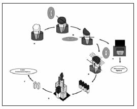
Fig. 1: Methodology used in the study. (A) Restorative Procedure. (B) Cutting the teeth into two sections. (C) Optic Microscopy Analysis (D) Transversal sections to obtain stick-shaped specimens for the microtensile test. (E) Microtensile test (F) Analysis of the fracture surface under stereoscopic microscope.
Microtensile Tests
The dental remainder was submitted to the microtensile bond strength tests, with its root end fixed to an acrylic block coupled to the metallographic cutter ISOMET 1000 (Buehler, Lake Bluff, IL, USA), which operated under water cooling at a speed of 800 rpm and static load of 80 grams. Sections were cut in planes across the plane of the dentin adhesive/resin composite interface, in the vestibular-lingual and mesio-distal directions of the tooth (Fig. 1).
The sticks obtained were fixed to the Universal Test Machine Instron Model 4411 (Instron Inc., Canton, MA, USA), with cyanoacrylate adhesive (Super Bonder - Henkel Ltda., Itapevi, Sao Paulo, Brazil) and subject to traction with a 50N load cell at a speed of 0.5 mm/min until they ruptured. The fractured area of each specimen was measured with a digital pachymeter (Digimess, Shinko Precision Gaging, Ind. Com. Ltda., China) to calculate the bond strength.
Data Analysis
The results obtained were submitted to Pearson's Correlation test, at a level of significance of α=0.05, to verify whether there was correlation between the resin tag lengths, hybrid layer thickness and the bond strength of the system studied.
Fracture Pattern Analysis
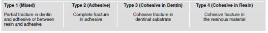
After the microtensile test, the fracture patterns were analyzed with the aid of a stereoscopic microscope Stemi SV 11 (Carl Zeiss Company - DSM-940 A, Oberkochen, Baden-Wurttemberg, Germany) at 40× magnification. To reveal the fracture area of each specimen more clearly, the staining solution proposed by Ohkubo et al., in 1982 19 was applied to the region corresponding to the bond interface, which stained the resin-free dental structure. The criteria proposed by Figueiredo Reis et al. 20 were used to analyze the fracture patterns (Table 3).
Table 3: Criteria for Fracture Pattern Analysis (Figueiredo Reis et al., 2003 20 with modifications).
RESULTS
The microscopy analysis of the specimens showed the presence of resin tags with a mean length of 4.34 μm (0.28) and mean hybrid layer thickness of 2.19 μm (0.34), this being uniform and continuous, and present throughout the full extent of the cut (Fig. 2); the microtensile test showed a mean value of 9.73 MPa (5.55). Application of Pearson's Correlation test, between the means of the factors microtensile bond strength and hybrid layer thickness (r=0.43) and between those of microtensile bond strength and resin tag length (r=0.014), showed absence of statistically significant correlation between the factors (Figs. 3 and 4).
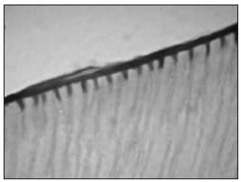
Fig. 2: Resin tags and hybrid layer observed by optical microscopy (Group II – E1) (1000× magnification).
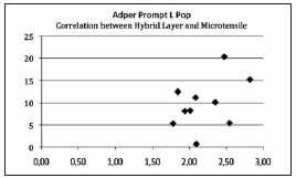
Fig. 3: Correlation test between hybrid layer and microtensile bond strength for the adhesive system Adper Prompt L Pop.
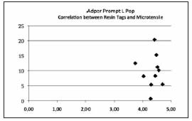
Fig. 4: Correlation test between resin tags and microtensile bond strength for the adhesive system Adper Prompt L Pop.
As regards fracture pattern, 68% adhesive fracture, 26% mixed fractures and 6% cohesive fractures in resin were observed, no cohesive fracture in dentin being found (95% confidence) (Fig. 5).
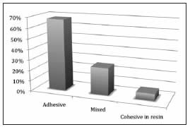
Fig. 5: Fracture patterns for the adhesive system Adper Prompt L Pop.
DISCUSSION
The excellent Brown & Brenn21 staining method and common optical microscopy clearly showed the resinous structures in a large area of the bond interface of the decalcified dentinal tissue12,13,18,22-24 in a single specimen. The mean values of hybrid layer thickness, resin tag length and microtensile bond strength of 2.19 μm, 4.34 μm and 9.73 MPa, respectively, obtained in the present "in vivo" study (Table 2) were lower than those from other studies that used the same adhesive system "in vitro"13,23.
It should be pointed out that with the aim of making the results more compatible with real-life situations, in this study the adhesive system was applied "in vivo", which may have contributed to these results, since the internal pressure in the dentinal tubules certainly diminishes the penetration of the adhesive system into the etched dentinal tissue25,26. Studies report that dentinal fluid pressure may cause lower bond strength values than those obtained in situations in which there is no pulp pressure present27-31. The low microtensile bond strength values obtained for the self-etching adhesive system used may also be related to the specimen storage conditions, in which the presence of moisture may have led to a degradation of the surface30,31 due to the considerable permeability of the bond interface in this material 30. In fact, Sauro et al., 200725 reported that the dentinal sealing desired after application of the adhesive system does not occur effectively in the self-etching systems due to the combination of the acid hydrophilic and hydrophobic monomers present in a single compartment, thus compromising the action of each of these components30,32. In addition to these considerations, the application of the one-step self-etching adhesive system could lead to bond failures as a result of the excess water present in its composition, which results in incomplete polymerization of the adhesive system. Therefore, water sorption by hydrophilic resinous monomers, both in the hybrid layer and in the tags, contributes to low tensile bond strength values25,32. Indeed, the images obtained by common optical microscopy (1000×) showed a weak bond of this adhesive to dentin (Fig. 3), with the more frequent occurrence of adhesive fractures.
In addition to the abovementioned factors, it should be noted that the low pH found in the non-polymerized surface areas of this adhesive system, due to inhibition by oxygen, could result in a reduction in tertiary amines, thereby leading to poor polymerization of the adhesive material, and consequently, a reduction in bond strength between the materials could occur more easily25. Furthermore, the low concentration of hydrophobic components at the bond/dentin interface could be responsible for the low bond strength values, because they provide lower hydrolytic resistance16,23,31. Certainly, these factors contributed to failures at the interface between the resin composite and adhesive system32,33, thus limiting the clinical success of the restoration. As in the "in vitro" research conducted by Oliveira et al., in 200923, this study found no correlation between the values for the hybrid layer and resin tag length and the microtensile bond strength of the self-etching adhesive system Adper Prompt L Pop to dentinal tissue, when it was applied "in vivo". This demonstrated that for this adhesive system, there may be no correlation between the variables under study. The non-existence of correlation between the hybrid layer thickness and microtensile bond strength may also be related to the intrinsic mechanical properties of each material, such as the degree of conversion and concentration of hydrophilic monomers8,11,32. The absence of correlation observed may be related to the findings of Spencer & Wang, in 200232, who reported that the application of this same system promoted profound migration of low molecular weight molecules, such as hydrophilic monomers (HEMA).
Since the greater part of the resin tags and the base of the hybrid layer are formed by low molecular weight monomers, being weakly polymerized therefore reduces their contribution to bond strength4. Similarly, Van Landuyt et al., in 200733 reported that for this system, the thickness of the hybrid layer formed and length of tags did not appear to be the main factors responsible for an effective bond; but the quality of the dentin/bond interface is the main factor to consider in self-etching adhesive systems, as it is determined by the components that form the chemical composition of the adhesive. The null hypothesis was accepted, since the microtensile bond strength of a self-etching adhesive system Adper Prompt L Pop did not depend on the thickness of the hybrid layer length of resin tags formed in healthy dentin in vivo.
CONCLUSION
Based on the results obtained, it may be concluded that the microtensile bond strength of a self-etching adhesive system Adper Prompt L Pop was not dependent on the thickness of the hybrid layer and length of resin tags.
1. Buonocore MG. A simple method of increasing the adhesion of acrylic filling materials to enamel surfaces. J Dent Res 1955;34:849-853. [ Links ]
2. Sattabanasuk V, Vachiramon V, Qian F, Armstrong SR. Resin-dentin bond strength as related to different surface preparation methods. J Dent 2007;35:467-475. [ Links ]
3. Nakabayashi N. Hybridization of natural tissues containing collagen with biocompatible materials: adhesion to tooth substrates. J Biomed Mater Res 1989;23:265-273. [ Links ]
4. Rocha PI, Borges AB, Rodrigues JR, Arrais CA, Giannini M. Effect of dentinal surface preparation on bond strength of self-etching adhesive systems. Braz Oral Res 2006; 20:52-58. [ Links ]
5. Chaves P, Giannini M, Ambrosano GM. Influence of smear layer pretreatments on bond strength to dentin. J Adhes Dent 2002;4:191-196. [ Links ]
6. Yesilyurt C, Bulucu B. Bond strength of total-etch dentin adhesive systems on peripheral and central dentinal tissue: a microtensile bond strength test. J Contemp Dent Pract 2006;7:26-36. [ Links ]
7. Pereira PN, Nunes MF, Miguez PA, Swift EJ Jr. Bond strengths of a 1-step self-etching system to caries-affected and normal dentin. Oper Dent 2006;31:677-681. [ Links ]
8. Walker MP, Wang Y, Spencer P. Morphological and chemical characterization of the dentin/resin cement interface produced with a self-etching primer. J Adhes Dent 2002; 4:181-189. [ Links ]
9. Tay FR, Pashley DH. Aggressiveness of contemporary selfetching systems I:. Depth of penetration beyond dentin smear layers. Dent Mater 2001;17:296-308. [ Links ]
10. Van Landuyt KL, Peumans M, De Munck J, Lambrechts P, Van Meerbeek B. Extension of a one-step self-etch adhesive into a multi-step adhesive. Dent Mater 2006;22:533-544. [ Links ]
11. Reis A, Grandi V, Carlotto L, Bortoli G, Patzlaff R, Rodrigues Accorinte Mde L, Dourado Loguercio A. Effect of smear layer thickness and acidity of self-etching solutions on early and long-term bond strength to dentin. J Dent 2005;33:549-559. [ Links ]
12. Tay FR, Gwinnett AJ, Pang KM, Wei, SHI. Micromorphologic relationship of the resin-dentin interface following a total-etch technique in vivo using a dentinal bonding system. Quintessence Int 1995a; 26:63-70. [ Links ]
13. Sundfeld RH, Valentino TA, de Alexandre RS, Briso AL, Sundefeld ML. Hybrid layer thickness and resin tag length of a self-etching adhesive bonded to sound dentin. J Dent 2005;33:675-681. [ Links ]
14. Sano H, Shono T, Sonoda H, Takatsu T, Ciucchi B, Carvalho R, Pashley DH. Relationship between surface area for adhesion and tensile bond strength-evaluation of a microtensile bond test. Dent Mater 1994;10:236-240. [ Links ]
15. Chen LJ, Meng QF, Chen YM, Smales RJ, Yip KH. Effect of fluoride iontophoresis on the microtensile bond strength between dentin and two adhesive systems. J Dent 2008; 36:697-702. [ Links ]
16. Brackett WW, Tay FR, Looney SW, Ito S, Haisch LD, Pashley DH. Microtensile dentin and enamel bond strengths of recent self-etching resins. Oper Dent 2008;33:89-95. [ Links ]
17. Pioch T, Stotz S, Buff E, Duschner H, Staehle HJ. Influence of different etching times on hybrid layer formation and tensile bond strength. Am J Dent 1998;11:202-206. [ Links ]
18. Sundfeld RH, Mauro SJ, Sundefeld MLMM, Briso ALF. Avaliacao clinico/microscopica da camada hibrida de adesao e dos prolongamentos resinosos (tags), em tecido dentinario condicionado. Efeitos de materiais, tecnicas de aplicacao e de analise. J Bras Dent Estet 2002;1:315-331. [ Links ]
19. Ohkubo N, Iwata S, Chikada K, Kuriyama S, Narita M, Ishikawa T, Yoshida T. A retention comparison of two sealants. Bull Tokyo Dent Coll 1982;23:201-219. [ Links ]
20. Figueiredo Reis A, Giannini M, Ambrosano GM, Chan DCN. The effects of filling techniques and a low-viscosity composite liner on bond strength to class II cavities. J Dent 2003;31:59-66. [ Links ]
21. Brown JH, Brenn L. A method for differential staining of Gram positive and Gram negative bacteria in tissue reactions. Bull Johns Hopkins Hospital 1931;48:69-73. [ Links ]
22. Sundfeld RH, da Silva AM, Croll TP, de Oliveira CH, Briso AL, de Alexandre RS, Sundefeld ML. The effect of temperature on self-etching adhesive penetration. Compend Contin Educ Dent 2006;27:552-556. [ Links ]
23. Oliveira FG, Anchieta RB, Rahal V, de Alexandre RS, Machado LS, Sundefeld ML, Giannini M, Sundfeld RH. Correlation of the hybrid layer thickness and resin tags length with the bond strength of a self-etching adhesive system. Acta Odontol Latinoam 2009;22:177-181. [ Links ]
24. Anchieta RB, Rocha EP, Ching-Chang KO, Sundfeld RH, Martin Junior M, Archangelo CM. Localized mechanics of dentin self-etching adhesive system. J Appl Oral Sci 2007; 15:321-326. [ Links ]
25. Sauro S, Pashley DH, Montanari M, Chersoni S, Carvalho RM, Toledano M, Osorio R, Tay FR, Prati C. Effect of simulated pulpal pressure on dentin permeability and adhesion of self-etch adhesives. Dent Mater 2007;23:705-713. [ Links ]
26. Hosaka K, Nakajima M, Monticelli F, Carrilho M, Yamauti M, Aksornmuang J, Nishitani Y, Tay FR, Pashley DH, Tagami J. Influence of hydrostatic pulpal pressure on the microtensile bond strength of all-in-one self-etching adhesives. J Adhes Dent 2007;9:437-442. [ Links ]
27. Ciucchi B, Bouillaguet S, Holz J, Pashley D. Dentinal fluid dynamics in human teeth, in vivo. J Endod 1995;21:191-194. [ Links ]
28. Campos EA, Correr GM, Leonardi DP, Barato-Filho F, Gonzaga CC, Zielak JC. Chlorhexidine diminishes the loss of bond strength over time under simulated pulpal pressure and thermo-mechanical stressing. J Dent 2009;37:108-114. [ Links ]
29. Vachiramon V, Vargas MA, Pashley DH, Tay FR, Geraldeli S, Qian F, Armstrong SR. Effects of oxalate on dentin bond after 3-month simulated pulpal pressure. J Dent 2008;36:178-185. [ Links ]
30. Sidhu SK, Omata Y, Tanaka T, Koshiro K, Spreafico D, Semeraro S, Mezzanzanica D, Sano H. Bonding characteristics of newly developed all-in-one adhesives. J Biomed Mater Res B Appl Biomater 2007;80:297-303. [ Links ]
31. Van Landuyt KL, Kanumilli P, De Munck J, Peumans M, Lambrechts P, Van Meerbeek B. Bond strength of a mild self-etch adhesive with and without prior acid-etching. J Dent 2006;34:77-85. [ Links ]
32. Spencer P, Wang Y. Adhesive phase separation at the dentin interface under wet bonding conditions. J Biomed Mater Res 2002;62:447-456. [ Links ]
33. Van Landuyt KL, Snauwaert J, De Munck J, Peumans M, Yoshida Y, Poitevin A, Coutinho E, Suzuki K, Lambrechts P, Van Meerbeek B. Systematic review of the chemical composition of contemporary dental adhesives. Biomaterials 2007;28:3757-3785. [ Links ]

