


Servicios Personalizados
Revista

Articulo
 Inglés (pdf)
 Articulo en XML
 Referencias del artículo
 Enviar articulo por email
Indicadores
-
 Citado por SciELO
Links relacionados
-
 Similares en
SciELO
Similares en
SciELO
Compartir

 Permalink
Acta Odontológica Latinoamericana
versión On-line ISSN 1852-4834
Acta odontol. latinoam. vol.25 no.3 Buenos Aires dic. 2012
ARTÍCULOS ORIGINALES
Can ultrasound application influence the bond strength of self-adhesive resin cements to dentin?
Diego F. F. da Silva1, Maurem L. Marcondes1, Niélli C. de Souza1, Bruna Gomes Daudt1, Luiz H. Burnett-Júnior1, Ana M. Spohr2
1 Department of Restorative Dentistry, School of Dentistry, Pontifical Catholic University of Rio Grande do Sul, Brazil.
2 Department of Dental Materials, School of Dentistry, Pontifical Catholic University of Rio Grande do Sul, Brazil.
CORRESPONDENCE Dr. Ana Maria Spohr PUCRS - School of Dentistry - block 6 Avenida Ipiranga, 6681 - CEP: 90619-900 Porto Alegre, RS, Brazil e-mail: ana.spohr@pucrs.br
ABSTRACT
The aim of this study was to evaluate the influence of ultrasound application on the bond strength of self-adhesives resin cements to dentin. Twenty-four third molars were randomly divided into 4 groups (n=6/group): G1 - Rely X Unicem; G2 – Maxcem Elite; G3 – RelyX Unicem and ultrasound application; G4 – Maxcem Elite and ultrasound application. Composite resin blocks were luted to flat dentin with a load of 500 g for 2 min, followed by light polymerization in G1 and G2. In G3 and G4, the ultrasound device was applied for 20 s on the composite resin block, followed by 500 g load for 1 min and 40 s, and light polymerization. After storage in distilled water at 37oC for 24 h, six tooth/resin sets were cut parallel to the long axis of the tooth, in the x and y directions, with a cross section area of ~0.80 mm2. Twenty-four specimens were obtained for each group and submitted to microtensile bond strength (μTBS) testing in a universal testing machine at 0.5 mm/min crosshead speed. According to two-way ANOVA, resin cement (p=0.000) and cementation method (p=0.002) were significant. Interaction was not significant (p=0.676). According to Student's-t test (α=0.05), the μTBS mean with ultrasound application (13.74 MPa) was statistically higher than without it (10.57 MPa). The μTBS mean of RelyX Unicem (13.95 MPa) was statistically higher than Maxcem Elite (10.36 MPa). The ultrasound application increased the μTBS of the RelyX Unicem and Maxcem Elite to dentin.
Key words: Dentin; Bond strength; Cements; Ultrasound.
RESUMO
A aplicação do ultrassom pode influenciar a resistência de união de cimentos resinosos autoadesivos à dentina?
O objetivo desse estudo foi avaliar a influencia da vibracao ultrassonica na resistencia de uniao de cimentos resinosos autoadesivos a dentina. Vinte e quatro dentes terceiros mo lares foram divididos aleatoriamente em quatro grupos (n=6/grupo): G1 – RelyX Unicem; G2 – Maxcem Elite; G3 – RelyX Unicem com vibracao ultrassonica; G4 – Maxcem Elite com vibracao ultrassonica. Blocos de resina composta foram cimentados sobre dentina plana com carga de 500 g por 2 min, seguido de fotoativacao nos G1 e G2. Em G3 e G4, a vibracao ultrassonica foi aplicada por 20 s sobre o bloco de resina composta, seguido de carga de 500 g por 1 min e 40 s e fotoativacao. Apos armazenagem em agua destilada a 37o C por 24 h, seis conjuntos dente/resina foram cortados paralelamente ao longo eixo do dente, nos sentidos x e y, com seccao de aproximadamente 0,8 mm2. Foram obtidos 24 corpos de prova para cada grupo, sendo entao submetidos ao teste de resistencia a microtracao (Rμt) em maquina de ensaio universal com velocidade de 0,5 mm/min. De acordo com Analise de Variancia Fatorial, as variaveis tipo de cimento resinoso (p=0,000) e tipo de cimentacao (p=0,002) foram significativas. A interacao das duas varaveis nao foi significativa (p=0,676). De acordo com o teste t-student (α=0,05) a media de Rμt com uso da vibracao ultrassonica (13,74 MPa) foi estatisticamente superior sem aplicacao da vibracao ultrassonica (10,57 MPa)). A media de Rμt do RelyX Unicem (13,95 MPa) foi estatisticamente superior ao Maxcem Elite (10,36 MPa). A vibracao ultrassonica aumentou a resistencia de uniao dos cimentos resinosos autoadesivos RelyX Unicem e Maxcem Elite a dentina.
Palavras-chave: Dentina; Resistencia de uniao; Cimentos; Ultrassom.
INTRODUCTION
Resin cements are widely used because their mechanical properties, aesthetics and ability to bond to restorative materials when proper pretreatment is applied are better than those of conventional cements1. A new category of resin cements – self-adhesive resin cements – have gained popularity with clinicians because they are easy to use and the luting procedure takes less time than with resin cements that require the application of an adhesive system. Without the adhesive system, part of the sensitivity of the technique is eliminated1,2. Despite being easier to apply, it is important that these selfadhesive materials should be capable of bonding adequately to both the dental structures and the restorative material in order to strengthen the tooth. Self-adhesive resin cements interact superficially with tooth hard tissues3,4. They have lower bond strength with enamel than resin cements requiring an adhesive system do2,5. In relation to dentin, some studies have shown that self-adhesive resin cements perform comparably to multi-step systems on coronal dentin2,3,5-7, although others have shown that they have significantly lower bond strengths to dentin8-10. To improve the bond, enamel etching with phosphoric acid has been suggested7,11; however, on dentin, this etching harms the effectiveness of the bond, which may be due to inadequate resin cement infiltration into the collagen fiber network3. With the aim of increasing the bond strength of selfadhesive resin cements to dentin, the application of weak acids, such as polyacrylic acid before the luting procedure has been tested12,13. Polyacrylic acid partially removes the smear layer14, leaves the mineral phase of dentin and increases the chemical reaction between the material and substrate15. However, it would be interesting to test other techniques, such as ultrasound application, with the aim of improving the adhesion of self-adhesive resin cements to dental substrate.
Studies have shown that ultrasound application during cementation affects the thixotropic properties of luting agents, leading to a decrease in viscosity16,17. This may promote an adequate wetting and adaptation of the densely filled resin cements to the dental substrate18. Ultrasound vibration reduced porosities in glass ionomer cements19, increased the temperature of the cement, shortened the setting reaction, and increased the bond strength to enamel20. The purpose of this study was to evaluate in vitro the effect of ultrasound application during the luting procedure on the bond strength of self-adhesive resin cements to dentin. This study was conducted under the null hypothesis that ultrasound application does not influence the bond strength of self-adhesive resin cements to dentin.
MATERIALS AND METHODS
Twenty-four unerupted human third molars, extracted for therapeutic reasons, were cleaned of gross debris and stored in distilled water at 4oC. The water was changed every week and the teeth were used within a period not exceeding 6 months. Roots were mounted in self-cured acrylic resin, and the occlusal enamel surface was removed with a low concentration diamond disc mounted on a lowspeed laboratory cutting machine Labcut 1010 (Extec Corp., London, England), under cooling. The rest of enamel was removed with 400 grit silicon carbide abrasive paper in a polishing machine DPU-10 (Panambra, Sao Paulo, SP, Brazil) under water. The superficial dentin was exposed and finished with 600 grit silicon carbide abrasive paper in the polishing machine, and a flat dentin surface was obtained. After polishing, the teeth were randomly divided into four groups (n=6) according to the materials used (Table 1) and treatment applied.
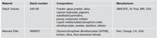
Table 1: Chemical composition and batch number of the resin cements.
Before the luting procedure, composite resin blocks of Z250 (3M, St. Paul, MN, USA) were obtained using a stainless steel mold with an inner diameter of 10 mm and a height of 5 mm. Three equal increments were inserted into the mold and each increment was light-cured for 40 seconds with a quartz-tungstenhalogen curing unit (XL 3000, 3M, St. Paul, MN, EUA). The surface of the resin composite block was treated with airborne particle abrasion with 50-μm aluminum oxide for 5 s at 4-bar pressure, and a layer of silane (Angelus, Londrina, PR, Brasil) and a layer of bond (Adhese, Ivoclar Vivadent, Liechtenstein) were applied, followed by light curing for 10 seconds.
Group 1 - RelyX Unicem: equal quantities of base and catalyst pastes were mixed and applied on dentin at approximately 1 mm thickness, and the composite resin block was luted to the tooth under a 500-g load by means of a metallic tool for 2 min. The excess resin cement was removed, followed by light curing for 40 s on each side (mesial, distal, buccal, lingual and occlusal) with the curing unit XL 3000. The light intensity was controlled by a radiometer (model 100, Demetron/Kerr, Danbury, CT, USA) in the interval between 450 and 500 mW/cm2. The specimens were stored for 24 h at 37°C in distilled water.
Group 2 - Maxcem Elite: the material was applied on dentin at approximately 1 mm thickness using the syringe supplied by the manufacturer, followed by the composite resin block as described for group 1.
Group 3 - RelyX Unicem with ultrasound application: the cement manipulation was the same as described for group 1. The composite resin block was luted under ultrasound vibration. An ultrasound tip provided with a rubber cap (C20 tip applicator, Gnatus, Ribeirao Preto, SP, Brazil) was mounted on an ultrasound handpiece (Jet Sonic Four Plus, Gnatus, Ribeirao Preto, SP, Brail), set at 30% of the power according to the manufacturer's recommendation. The operator held the handpiece and the tip was oriented perpendicular to the surface of the composite resin block. The vibration was applied for 20 seconds, in an intermittent mode, under a load of approximately 100g. This load was previously calibrated by the operator using a digital weighing-machine. After the ultrasound application, the 500-g load was applied for 1 min and 40 seconds. The excess resin cement was removed, followed by light-curing as described for group 1.
Group 4 - Maxcem Elite with ultrasound application: the cement manipulation was the same as described for group 2. The luting procedure was the same as described for group 3.
Specimens were stored for 24 h at 37oC in distilled water, and then sectioned perpendicular to the bonding surface using a Labcut 1010 laboratory cutting machine at a speed of 400 rpm with a diamond disk under water cooling. The specimens had a cross-section of approximately 0.90 × 0.90 mm, measured with a digital caliper (Mitutoyo Sul Americana Ltda., Suzano, SP, Brazil). Six sticks from the central region of each tooth were used, which were examined with a stereomicroscope (Olympus Corp., Tokyo, Japan) at 25× magnification to analyze the adhesive area. The specimens with defects such as bubbles, lack of material or irregular areas were discarded. Twentyfour specimens were selected for each group. The specimens were then fitted to the microtensile testing device, which has two stainless steel grips with an area of 8 × 10 mm and sliding shafts that prevent torsion movements during the tests. These shafts have a fixing screw that prevents the specimen from moving during bonding. The specimens were fixed with cyanoacrylate glue (Loctite, Sao Paulo, SP, Brazil), associated with the Zip Kicker accelerator (Pacer Technology, Rancho Cucamonga, CA, USA), and stressed at a crosshead speed of 0.5 mm/min until failure in a universal testing machine (EMIC DL-2000, Sao Jose dos Pinhais, PR, Brazil) using a cell load of 50 N. The μTBS was expressed in MPa and derived by dividing the force (N) at the time of fracture by the bond area (mm2). The fractured surfaces of all specimens were observed by scanning electron microscopy (SEM) (Philips XL 30, Philips Electronic Instruments Inc., Mahwah, NJ, USA). The failures were classified as adhesive (failure between dentin and resin cement), cohesive in dentin (dental substrate failure), cohesive in resin cement (failure inside the resin cement) or mixed (two or more types of failure). The specimens were further analyzed regarding the percentage of remaining resin cement on the dentin surface. Images of the fractured areas from SEM at X200 magnification were viewed on a 15-inch computer screen. A grid divided into 16 squares was placed over the specimen image (Fig. 1). The criterion used to classify a "square with cement" was the presence of remaining cement in at least half of the square. The remaining cement area on the fractured surface of each specimen was calculated as percentage of the total area. Data on the percentage of remaining cement for each specimen were recorded, and the mean values for each surface treatment were calculated.
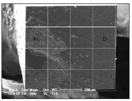
Fig. 1: Dentin μTBS fracture surfaces of a representative sample treated with RelyX Unicem showing mixed failure. RC: resin cement; D: dentin.
Two-way analysis of variance (ANOVA) was used to test the effect of the resin cements and the cementation method on the μTBS. Furthermore, Student t-test was used to determine differences in μTBS between the resin cements, and with and without ultrasound application. P<0.05 was considered significant. The software used was SPSS 10.0 (SPSS Inc., Chicago, IL, USA).
RESULTS
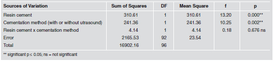
Two-way ANOVA analysis showed that the resin cement (p=0.000) and the cementation method (p=0.002) had a significant effect on the μTBS, while the interaction effect was not significant (p=0.676) (Table 2).
Table 2: Results for the two-way ANOVA.
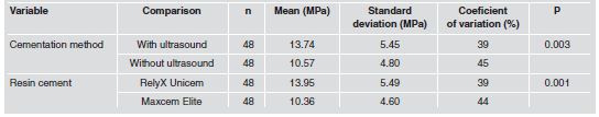
According to Student-t test, the μTBS mean with ultrasound application (13.74 MPa) was statistically higher than without ultrasound application (10.57 MPa) (p=0.003). The μTBS mean for RelyX Unicem (13.95 MPa) was statistically higher than Maxcem Elite (10.36 MPa) (p=0.001) (Table 3).
Table 3: μTBS for the experimental groups.
All specimens showed mixed failure (adhesive + cohesive in resin cement). The percentage values of remaining resin cement on the dentin surface was 16.8% for group 1, 25% for group 2, 15.3 % for group 3 and 19.2% for group 4.
DISCUSSION
The null hypothesis was rejected, because ultrasound application increased the μTBS of RelyX Unicem and Maxcem Elite self-adhesive resin cements to dentin. In this study, ultrasound vibration was applied for 20 s during the cementation procedure of the composite resin block, allowing the resin cement to flow and the resin block to become seated on the dentin. After this, a static load of 500g was applied. Although 20 s correspond to a rapid procedure, they were sufficient to influence the μTBS value. Whenever two materials are mixed, whether they are powder and liquid, or a paste – paste system, air bubbles are incorporated, making the material more porous. This porosity reduces the intrinsic strength of the material21, and may reduce its bond capacity to some substrate. Cantoro et al.22 used SEM to verify that ultrasound vibration reduced the porosity of the self-adhesive resin cements RelyX Unicem and G-Cem, making them more homogeneous. The same effect was observed on glass ionomer cements19. The reduction in resin cement porosity might have allowed these materials to wet the dentinal substrate better, promoting a higher mean μTBS when cementation was associated with ultrasound application.
It has been reported that ultrasound vibration increases the mechanical properties of glass ionomer cements, especially in the first 24 h23, as well as the bond strength to dentin24. Algera et al.20 studied resin-modified glass ionomer cements, reporting that the local heat produced by ultrasound vibration may catalyze free radicals during polymerization, favoring improvement in the mechanical properties and a better bond to the substrate. As regards self-adhesive resin cements, Cantoro et al.22 reported that ultrasound vibration could also favor a greater initial reaction between the calcium hydroxide and acid resin monomers of RelyX Unicem, a reaction in which the water required for ionization of the functional monomers is generated. In addition, the acid-base reaction between the acid monomers and basic inorganic particles of the material must also be increased22. It is therefore suggested that ultrasound application favored a greater setting reaction in both the self-adhesive resin cements studied, providing an increase in the mechanical properties in the first 24 h, and thus, greater bond strength. Another important effect of ultrasound vibration is on the thixotropic properties of cementing agents. It was demonstrated that oscillatory load on the cement increased its fluidity, leading to a reduction in viscosity17. De Munck et al.3 observed high viscosity and presence of spaces in the resin cement layer for RelyX Unicem, resulting in insufficient adaptation to dental substrate. It is therefore suggested that when the ultrasound vibration reached the resin cements RelyX Unicem and Maxcem Elite, it caused an intrinsic vibration of the molecules and particles of the cements precisely at the moment at which the shear forces were being placed on the material by the cementation load. This favored a reduction in viscosity of the resin cement, greater wetting capacity and adaptation to the dentin, as well as a higher bond strength value, since the chemical and physical interactions with dentin are favored by the greater adaptation between the cement and substrate.
Regardless of the resin cement and whether or not ultrasound was applied, all specimens showed mixed failures, characterized by rupture at the level of the dentin – resin cement interface (adhesive), with the presence of cohesive failure in the resin cement itself, resulting in a certain quantity of resin cement remaining adhered to the dentin after the microtensile bond strength test. This type of failure demonstrates that the bond interface is the most fragile site, and is susceptible to rupture. However, a certain bond exists between self-adhesive resin cements and dentin, because part of the resin cement remained bond to the substrate. It was also observed that the presence of remaining cement was similar between the experimental groups. RelyX Unicem had higher μTBS (13.95 MPa) than Maxcem Elite (10.36 MPa), in agreement with other studies5,9. In general, self-adhesive resin cements have a limited capacity to demineralize tooth hard tissues3-5. The following hypotheses might explain these findings: (1) the pH of these cements, which is approximately 2.14, is not low enough; (2) the high viscosity of the cement3, and (3) neutralization may occur during mixture due to the chemical reaction that releases water or alkaline particles, which may increase the pH4. Studies evaluating the bond interface of self-adhesive resin cement using SEM 3,4,6 and TEM 3,25 showed no formation of a hybrid layer or resin tags. The bonding mechanism of RelyX Unicem to dentin appears to be chemical rather than micromechanical in nature26. This bond is established by the specific multifunctional phosphoric-acid methacrylates, which are ionized at the time of mixing and which react with the hydroxyapatite of the mineral tissues of the tooth26. According to information from the manufacturer, Maxcem Elite also contains an acid monomer, glycerol dimethacrylate dihydrogen phosphate (GPDM), which is partly responsible for the effect of etching and adhesion to the dental structure (Technical Bulletin, 2007).
Various factors may influence the bonding capacity and adhesion of resin cements to dentin, including chemical composition, viscosity and pH. Maxcem Elite tends to maintain its low pH (2.2), while the pH of RelyX Unicem increases after 48 h (from 2.8 to 7.0). Although low pH is necessary for adequate dentin etching, it has been speculated that if the pH is maintained for a long time, as in the case of Maxcem Elite, there could be an adverse effect on the bond between this cement and the dental structure27. Therefore, this characteristic of Maxcem Elite pH could be one of the factors that led to this material having lower bond strength than RelyX Unicem. The mean difference in μTBS with and without the ultrasonic application was 3 MPa. Although this difference is statistically significant, it is doubtful whether it would be of any clinical significance. To date, the literature has not established the minimum bond strength required between the material and the substrate to guarantee the success and longevity of the cementation procedure. Nevertheless, this study demonstrated that ultrasound may be applied without producing a negative effect on the process of bonding to dentinal substrate, but rather, a positive effect. Moreover, cementation with ultrasound application results in the restoration adapting to the preparation more rapidly, requiring a lower cementation load to seat the restoration in comparison with a static load, and a thinner cementation line18.
According to the methodology used and within the limitations of this study, the results suggest that the ultrasound application increased the μTBS of the self-adhesive resin cements RelyX Unicem and Maxcem Elite to dentin.
1. Burke FJ. Trends in indirect dentistry: 3. Luting materials. Dent Update 2005;32:257-260. [ Links ]
2. Abo-Hamar SE, Hiller KA, Jung H, Federlin M, Friedl KH, Schmalz G. Bond strength of a new universal self-adhesive resin luting cement to dentin and enamel. Clin Oral Investig 2005;9:161-167. [ Links ]
3. De Munck J, Vargas M, Van Landuyt K, Hikita K, Lambrechts P, Van Meerbeek B. Bonding of auto-adhesive luting material to enamel and dentin. Dent Mater 2004; 20:963-971. [ Links ]
4. Monticelli F, Osorio R, Mazzitelli C, Ferrari M, Toledano M. Limited decalcification/diffusion of self-adhesive cements into dentin. J Dent Res 2008;87:974-979. [ Links ]
5. Goracci C, Cury AH, Cantoro A, Papacchini F, Tay FR, Ferrari M. Microtensile bond strenght and interfacial properties of self-etching and self-adhesive resin cements used to lute composite onlays under different seating forces. J Adhes Dent 2006;8:327-335. [ Links ]
6. Al-Assaf K, Chakmakchi M, Palaghias G, Karanika-Kouma A, Eliades G. Interfacial characteristics of adhesive luting resins and composites with dentine. Dent Mater 2007;23: 829-839. [ Links ]
7. Hikita K, Van Meerbeek B, De Munck J, Ikeda T, Van Landuyt K, Maida T, Lambrechts P, Peumans M. Bonding effectiveness of adhesive luting agents to enamel and dentin. Dent Mater 2007;23:71-80. [ Links ]
8. De Angelis F, Minnoni A, Vitalone LM, Carluccio F, Vadini M, Paolantonio M, D'Arcangelo C. Bond strength evaluation of three self-adhesive luting systems used for cementing composite and porcelain. Oper Dent 2011;36: 626-634. [ Links ]
9. Lurs AK, Guhr S, Gunay H, Geurtsen W. Shear bond strength of self-adhesive resins compared to resin cements with etch and rinse adhesives to enamel and dentin in vitro. Clin Oral Investig 2010;14:193-199. [ Links ]
10. Viotti RG, Kasaz A, Pena CE, Alexandre RS, Arrais CA, Reis AF. Microtensile bond strength of new self-adhesive luting agents and conventional multistep systems. J Prosthet Dent 2009;102:306-312. [ Links ]
11. Duarte S Jr, Botta AC, Meire M, Sadan A. Microtensile bond strengths and scanning electron microscopic evaluation of self-adhesive and self-etch resin cements to intact and etched enamel. J Prosthet Dent 2008;100: 203-210. [ Links ]
12. Pavan S, dos Santos PH, Berger S, Bedran-Russo AK. The effect of dentin pretreatment on the microtensile bond strength of self-adhesive resin cements. J Prosthet Dent 2010;104:258-264. [ Links ]
13. Tonial D, Ghiggi PC, Lise AA, Burnett-Junior LH, Oshima HMS, Spohr AM. Effect of conditioner on microtensile bond strength of self-adhesive resin cements to dentin. Stomatologija 2010;12:73-79. [ Links ]
14. Araujo MAJ, Rode SM, Villela LC, Goncalves RD. Qualitative evaluation of the effect of cleaning agent on smear layer: structural study using scanning electron microscopy. Rev Odontol USP 1998;12:99-104. [ Links ]
15. Inoue S, Van Meerbeek B, Abe Y, Yoshida Y, Lambrechts P, Vanherle G, Sano H. Effect of remaining dentin thickness and the use of conditioner on micro-tensile bond strength of glass-ionomer adhesive. Dent Mater 2001;17: 445-455. [ Links ]
16. Judge RB, Wilson PR. The effect of oscillating force upon the flow of dental cements. J Oral Rehabil 2001;26:892-899. [ Links ]
17. Yu Z, Strutz JM, Kipnis V, White SN. Effect of dynamic loading methods on cement film thickness in vitro. J Prosthodont 1995;4:251-255. [ Links ]
18. Schmidlin PR, Zehnder M, Mityko CS, Gohring TN. Interface evaluation after manual and ultrasonic insertion of standardized class I inlays using composite resin materials of different viscosity. Acta Odontol Scand 2005;63:205-212. [ Links ]
19. Coldebella CR, Santos-Pinto L, Zuanon ACC. Effect of ultrasonic excitation on the porosity of glass ionomer cement: A scanning electron microscope evaluation. Microscop Res Tech 2001;74:54-57. [ Links ]
20. Algera TJ, Kleverlaan CJ, de Gee AJ, Prahl-Anderson B, Feilzer AJ. The influence of accelerating the setting rate by ultrasound or heat on the bond strength of glass ionomers used as orthodontic bracket cements. Eur J Orthod 2005; 27:472-476. [ Links ]
21. Nomoto R, McCabe JF. Effect of mixing methods on the compressive strength of glass ionomer cements. J Dent 2001;29:205-210. [ Links ]
22. Cantoro A, Goracci C, Conoglio I, Magni E, Polimeni A, Ferrari M. Influence of ultrasound application on inlays luting with self-adhesive resin cements. Clin Oral Investig 2011;15:617-623. [ Links ]
23. Towler MR, Bushby AJ, Billington RW, Hill RG. A preliminary comparison of the mechanical properties of chemically cured and ultrasonically cured glass ionomer cements, using nano-indentation techniques. Biomaterials 2001;22: 1401-1406. [ Links ]
24. Fagundes TC, Barata TJE, Bresciani E, Cefaly DFG, Carvalho CAR, Navarro MFL. Influence of ultrasonic seating on tensile bond strength of glass-ionomer cement to dentin. J Adhes Dent 2006;8:401-407. [ Links ]
25. Yang B, Ludwig K, Adelung R, Kern M. Micro-tensile bond strength of three luting resins to human regional dentin. Dent Mater 2006;22:45-56. [ Links ]
26. Gerth HUV, Dammaschke T, Zuchner H, Schafer E. Chemical analysis and bonding reaction of RelyX Unicem and Bifix composites— a comparative study. Dent Mater 2006; 22:934-941.
27. Han L, Okamoto A, Fukushima M, Okiji T. Evaluation of physical properties and surface degradation of self adhesive resin cements. Dent Mater J 2007;26:906-914. [ Links ]

