


Servicios Personalizados
Revista

Articulo
 Inglés (pdf)
 Articulo en XML
 Referencias del artículo
 Enviar articulo por email
Indicadores
-
 Citado por SciELO
Links relacionados
-
 Similares en
SciELO
Similares en
SciELO
Compartir

 Permalink
Acta Odontológica Latinoamericana
versión On-line ISSN 1852-4834
Acta odontol. latinoam. vol.26 no.1 Buenos Aires abr. 2013
ARTÍCULOS ORIGINALES
Dental caries experience in school children and the impact of non-cavitated lesions on the caries index
Ana M. Acevedo1, Maglynert Montero1, Carolina Machado2, Ilse Sáez2, Fátima Rojas - Sánchez1, Israel Kleinberg3
1 Institute of Dental Research "Raul Vincentelli", School of Dentistry, Central University of Venezuela. Caracas, Venezuela.
2 Department of Dentistry, Sucre Municipality, Miranda State, Caracas, Venezuela.
3 Division of Translational Oral Biology. Department of Oral Biology and Pathology, Stony Brook University. New York, USA.
CORRESPONDENCE Dr. Maglynert Montero Baptista Universidad Central de Venezuela. Facultad de Odontologia, Instituto de Investigaciones Odontologicas "Raul Vincentelli", Piso 9, Ciudad Universitaria, Los Chaguaramos Caracas, Venezuela. E-mail: maglymontero@yahoo.com
ABSTRACT
The purpose of this study was to evaluate the current status of dental caries in 11 to 13 year-old schoolchildren residing in Sucre Municipality, Miranda State, and the impact of the noncavitated lesion on the caries index. Twelve elementary schools were included in this study and a sample of 1484 children was examined using artificial light, a #5 mirror and a #23 probe. The criteria followed were those proposed by Radike (1972) as modified by Acevedo et al. (2005) in order to include initial non-cavitated caries lesions. Teeth were cleaned and dried for 5 seconds with a triple syringe. Caries prevalence was 94.07% and the average DMFS index for the total sample was 4.35 } 4.21. This increased significantly to 6.45 } 5.01, when the initial caries lesions were included (p <0.05). According to gender, DMFS was higher in the female population (4.51 } 4.45) than in males (4.21 } 3.97), but the difference was not statistically different (p> 0.05). The same pattern was observed, when the initial caries lesions were added. The new mean DMFS was 6.67 } 5.15 and 6.26 } 4.88 for females and males, respectively. Non-cavitated lesions represent 33% of the total caries lesions recorded. Conclusion: These results show that (i) dental caries prevalence in this population remains high and (ii) initial lesions contribute significantly to the DMFS index.
Key words: Dental Caries; Prevalence; Children.
Experiencia de caries dental en niños escolarizados y el impacto de las lesiones no cavitadas en el índice de caries
RESUMEN
El objetivo de este estudio fue evaluar la experiencia de caries en escolares de 11 a 13 anos de edad del Municipio Sucre y el impacto de las lesiones no cavitadas en el indice de caries. Se selecciono una muestra a conveniencia de 12 escuelas ubicadas en el Municipio Sucre, Estado Miranda, Venezuela. Previo consentimiento informado, se examinaron 1484 ninos utilizando luz artificial, espejo #5 y explorador #23; siguiendo los criterios propuestos por Radike (1972), modificados por Acevedo et al (2005). La deteccion de las lesiones iniciales se realizo previa profilaxis y secado del diente durante 5 segundos (con jeringa triple). Resultados: La prevalencia de caries fue de 94,07% y el indice CPOS promedio fue de 4,35}4,21 aumentando significativamente a 6,45}5,01 cuando se incorporaron las lesiones iniciales (p<0,05). De acuerdo al genero, se observo un CPOS mayor en las ninas (4,51}4,45) al compararlo con los varones (4,21}3,97), sin diferencia estadisticamente significativa (p>0,05). El mismo patron se observo cuando se incorporaron las lesiones iniciales al CPOS, 6,67}5,15 para ninas y 6,26}4,88 para varones. Las lesiones no cavitadas representaron el 33% del total de lesiones cariosas. Conclusion: Los resultados indican una alta prevalencia de caries en la poblacion estudiada, asi como la importancia de incluir las lesiones no cavitadas al indice CPOS.
Palabras Clave: Caries; Prevalencia; Niños.
INTRODUCTION
Dental caries is a process that involves an imbalance of the dynamic and physiological activities of demineralization and re-mineralization in the dental surface due to intermittent acid attack. If the disease is not controlled over time, net mineral loss can occur, with subsequent cavity formation1. This mineral loss does not always lead to cavity formation; nevertheless, the signs of the disease should be recognized as a series of continuous changes in severity and not just as a simple cavity on the dental surface. The detection of caries lesions in the non-cavitated stages is extremely important even in epidemiological surveys, because the caries situation of a population can be more accurately described2 and in a shorter time.
The results of studies in Venezuela are similar to those in other parts of the world. Dental caries prevalence has decreased over time3, presumably in response to the implementation of massive prevention programs. Nevertheless; all of these studies report the caries lesion as a "cavity", using traditional criteria such as those suggested by the World Health Organization4. White, chalky-looking spots or areas, where a probe is retained in the absence of softened tissue are considered healthy. In 1987, using these criteria, caries prevalence was found to be 83% with a DMFT index of 3.67 in 12 year old children in Venezuela, (Fundacredesa 1981 - 1987). Ten years later, Rivera et al. (unpublished data), showed a reduction in the DMFT index (2.12 } 2.49) for the same age group, and more recently, Moron et al.5 found a mean DMF of 1.20 for the same group of children, indicating a reduction in the caries index, when compared to the results obtained by Rivera.
A cross-sectional study performed by Acevedo et al.6 in Sucre Municipality, Miranda State, using the DMFS index and detection criteria proposed by Radike7 showed a dental caries prevalence of 74% for 9 to 13 year-old schoolchildren, with a mean DMFS index of 3.64 } 0.29 for the 12 year-olds involved. In 2005 8, the same group of researchers evaluated which teeth were most affected by dental carries in a population between 10 . and 11 years of age, residing in Sucre Municipality. The results showed that the lower first permanent molars were affected more than the upper first molars and the occlusal followed by the buccal surface had the highest percentage of involvement.
It is evident that the use of more specific detection criteria provides more information, when used in epidemiological and clinical studies. The use of methods that evaluate the early stages of the lesion are determinant for establishing the actual situation of dental caries disease and the need for its use should be emphasized in epidemiological studies to be performed in Latin America. In a study by Alvarez et al. 9 on preschool children from Sucre Municipality, where non-cavitated caries lesions were included, the results showed dental caries prevalence of 79%, and there were fewer non-cavitated than cavitated lesions.
The purpose of this study was to evaluate dental caries experience in schoolchildren and the impact of non-cavitated lesions on the caries index in school children residing in Sucre Municipality, Miranda State, using detection criteria that include lesions in both stages of development
METHODS AND SUBJECTS
Most of the Venezuelan population lives in urban and only 20% in rural areas. Sucre Municipality is one of the most populated marginal urban areas in Miranda State and has a low socio-economic status.
Universe and study sample
A sample of twelve elementary schools (national, state and municipal) located in an area of low socio-economic level of the Sucre Municipality in Venezuela was selected. Twelve schools were selected in random order from a list of schools and numbered from 1 to 12. The study unit was selected in a non-probabilistic manner, and all children were 11 to 13 years of age. The final sample consisted of 1,484 children. The total population registered in elementary schools in Sucre Municipality was 13,637 children; the sample accordingly represented 10.9% of the total population. Prior to clinical examination, the parents and/or the legal representatives of the children were informed of the study objectives and were asked to sign an informed consent form which was endorsed by the research examiner.
Clinical Examination
The oral examination was performed by a previously calibrated examiner (CM, who showed an intra-examiner kappa value of 0.89). This was done in a mobile dental unit, using artificial light, a # 5 flat mirror and a # 23 probe, after completing a thorough cleaning of the dental surface with a small brush installed on a low-speed hand piece. The children evaluated received preventive dental treatment and educational talks that included the use of the toothbrush and fluoridated toothpastes. The detection of dental caries lesions was performed after a dental prophylaxis using the diagnostic criteria proposed by Radike and modified by Acevedo et al.8 which included non-cavitated lesions.
Statistical Analysis
The data was analyzed using the SPSS 12.0 program (SPSS Inc., Chicago, USA). The kappa value was used to determine the intra- and inter-examiner reproducibility. The mean DMFS values and standard deviations were calculated. Student's t Test was used to establish possible differences between the different variables.
RESULTS
The total sample was distributed according to gender among 782 male subjects (52.7%) and 702 female (47.3%) subjects. The children were distributed according to age as follows: 644 children were 11 years; 598 were 12 years and 242 were 13 years old (Table 1).
Table 1: Sample distribution by age and gender.

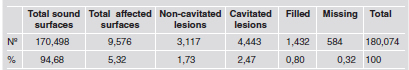
The results showed dental caries prevalence of 94.1%; total sound surfaces (94.7%) were considerably higher than affected surfaces (5.3%) (Table 2). The analysis of DMFS components showed that the carious component was prevalent (4.2%) and lesion distribution within this component was 2.5% cavitated lesions and 1.7% non-cavitated lesions. Filled surfaces represented 0.8%, and missing surfaces 0.3% (Table 2).
Table 2: Number and percentage of evaluated surfaces in each of the DMFS index components, in a sample of 1,484 school children between ages 11 and 13 in Sucre Municipality, Miranda State.
Diagnostic criteria proposed by Radike as modified by Acevedo et al.11
The mean DMFS index for the whole sample was 6.45 } 5.01. When the non-cavitated caries lesions were excluded, the mean index decreased significantly to 4.35 } 4.21 (p<0,001) (Table 3). The mean dmfs index according to age was 5.23}3.89 in the 11 year-old group; 6.79}5.26 in 12 year-olds; and 8.87}5.95 in 13 year-olds. When the non-cavitated caries lesions were excluded, the index decreased significantly (p<0.001) in each of the age groups 3.42}3.23; 4.63}4.44; and 6.13}5.15 for the 11, 12, and 13 year-olds, respectively. In both cases, the mean caries index increased proportionally to age (Table 3). According to gender, no statistically significant difference was observed between genders (Table 4), with mean DMFS values of 6.67 } 5.15 for females (4.51 } 4.45), when the non-cavitated lesions were excluded) and 6.26}4.88 for males (4.21 } 3.97) when the non-cavitated lesions were excluded.
Table 3: Contribution of the non-cavitated lesions to the DMFS index according to age, in a sample of 1,484 school children between ages 11 and 13 in Sucre Municipality, Miranda State.

Mean ± Standard Deviation of the DMFS index in a sample of 1,484 school children in Sucre Municipality, Miranda State. Nº = children evaluated per age. *A statistically significant difference was found between DMFS and DMFS excluding non-cavitated caries lesions (p<0.001) and the different ages (p<0.001).
Table 4: Contribution of the non-cavitated lesions to the DMFS index according to gender in a sample of 1,484 school children between ages 11 and 13 in Sucre Municipality, Miranda State.

Mean ± Standard Deviation of the DMFS index in a sample of 1,484 school children in Sucre Municipality, Miranda State. Nº = children evaluated per age. *A statistically significant difference was found between DMFS and DMFS excluding non-cavitated caries lesions (p<0.001). There was no difference in gender (p>0.001).
Finally, the percentage of sound surfaces and/or caries-affected surfaces was determined on each tooth (Table 5). The results showed that 13.6% of the occlusal surfaces were affected, followed by 7.7% of the buccal surfaces, 3.2% of the lingual, 1.1% of the mesial, and 0.9% of the distal surfaces. When all surfaces were considered, it is important to note that the total number of sound surfaces is considerably higher than the number of surfaces affected by caries (> 86%). When considering the type of lesion, non-cavitated caries lesion was the most prevalent on occlusal surfaces (5.68%), followed by cavitated caries lesions (5.13%). Comparing the percentage of cavitated and non-cavitated lesions on the buccal, lingual, mesial and distal surfaces, the percentage of cavitated lesions was higher than the percentage of non-cavitated lesions (p<0.001).
Table 5: Percentage of the different surfaces evaluated in each of the DMFS components in a sample of 1,484 school children between ages 11 and 13 in Sucre Municipality, Miranda State.

Diagnostic criteria proposed by Radike modified by Acevedo et al.8
Table 6 shows the contribution of non-cavitated caries lesions to the DMFS index. All surfaces except the distal show a significant reduction in the DMFS index when the non-cavitated lesions are excluded (p<0.001). The DMFS index for the occlusal surface was 3.32}2.80, followed by the buccal surface 1.87}1.28. The lingual, mesial, and distal surfaces had lower indexes: 0.78}1.12; 0.26}0.70; and 0.22}0.63, respectively.
Table 6: Contribution of the non-cavitated lesions to the DMFS index per dental surface in a sample of 1,484 school children between ages 11 and 13 in Sucre Municipality, Miranda State.

Mean ± Standard Deviation of the DMFS index in a sample of 1,484 school children in Sucre Municipality, Miranda State. *A statistically significant difference was found between DMFS and DMFS excluding non-cavitated caries lesions (p<0.001).
DISCUSSION
The results in this study show the status of dental caries in children residing in Sucre Municipality, Miranda State, Venezuela. It is important to note that these results cannot be extrapolated to other communities nationwide. Another aspect to consider is that the DMFS index used includes non-cavitated and cavitated caries lesions, which allows the examiner to make a more comprehensive assessment of the caries situation; nevertheless this makes it impossible to compare this study with other national and international studies where the DMFS index has been used, following other detection criteria.
The detection of caries lesions on the occlusal surfaces of the posterior teeth, particularly in pits and fissures and in its earliest stages, is a difficult task10-13, as is making a successful diagnosis of early lesions, especially those that are noncavitated14. The WHO reports the inaccuracy of lesion detection in stages previous to cavitations; therefore the results would not be reproducible15. In contrast, different studies have shown that inter-and intra-examiner reproducibility of observations is possible9. In this sense, our study showed a high intra-examiner kappa value (0.89), which indicates good reproduction of the observed data.
While dental caries prevalence has diminished significantly since massive preventive and restorative programs were implemented, it is also true that it has not been completely controlled. Data previously obtained in this Municipality showed that in the year 2000, dental caries prevalence in 9 to 13 year-olds was 74%, with a mean DMFS index of 3.64 } 0.29 at 12 years of age6; eight years later, this study has shown a dental caries prevalence of 94% with a mean DMFS index of 4.63 } 4.44 at 12 years of age (when non- cavitated caries lesions are not included). A comparison of the two studies shows that the number of subjects affected by caries increased by 27% and the index is 1.27 times higher. These figures suggest that all the effort made by the local authorities, such as implementation of massive educational programs, use of fluoridated tooth pastes, and the intake of fluoridated salt, which despite its implementation in 1995 does not meet the established standard (unpublished data) has not been enough to control the disease. It is also true that the social determinants such as low quality of life, inadequate eating patterns, sanitation services, drinking water, and deficient sewage, among others to which the population is exposed have not been taken into account16 and keep this population at risk of develop the disease. It is worth noting that in spite of the high caries prevalence observed in the study population, there was a low percentage of affected surfaces (5.32%), showing that the disease continues to be prevalent but only affects a few dental surfaces per individual. This is consistent with the study published by Larmas17, which reported that the real caries experience of a population must include not only the prevalence of the disease but also detailed data that can help further understanding of the disease.
The evaluation of different components of the DMFS index shows that the decay component was the most prevalent, and by using more updated detection criteria it was possible to differentiate cavitated and non-cavitated lesions, with cavitated lesions generally being the most prevalent (2.5%). On the other hand, when the occlusal surfaces were evaluated alone, a different pattern was observed, with non-cavitated lesions being significantly more prevalent (p>0.0001) than cavitated lesions, suggesting a delay of the disease on this surface. These results suggest that the use of these caries detection criteria makes it possible to register occlusal caries lesions at the earliest stages of progression. However, it is difficult to predict whether these lesions are at the enamel level or have reached the dentinenamel junction or even the dentin. Pits indicate that the detectable lesion represents a more advanced stage of the lesion18, however they can be reverted or their progression controlled with the bioavailability of calcium, phosphate and fluoride present in the biofilm, which favor the remineralization process. The filled component of the DMFS index represented a very low percentage, indicating the lack of restorative attention by dental health care institutions in this population.
The results of this study indicated that the occlusal surface was the most affected tooth caries site. These results are supported by previous research conducted in Sucre Municipality8 and by other papers published around the world19-21. The second most affected was the buccal surface, which makes a major contribution to the DMFS index due to the presence of the buccal groove, which favors plaque retention and therefore caries development. The proximal surfaces (mesial and distal) do not make a major contribution to the index, not because they are not susceptible to dental caries, but in epidemiological studies they are under-recorded because of the limited access for caries detection. Different authors therefore suggest the use of different techniques to obtain better results, such as elective temporary separation (to observe proximal caries), drying the dental surface, and the use of x-rays22. The truth is, that in countries where attention and prevention resources are as limited as ours, the use of other diagnosis methods such as DIAGNOdent, FOTI (Fiber Optic Transilumination) and QLF (Quantitative Light-Induced Fluorescence)23 involves higher costs, so the excellent sensitivity they provide must be left aside in favor of the low sensitivity and adequate specificity provided by the visual tactile method. A viable alternative is the use of more specific detection criteria to detect lesions in all progression stages. Currently, ICDAS (International Caries Detection and Assessment System) developed by a consensus Cariology group shows great diagnostic potential when compared to traditional methods24. However, although it allows evaluation of the progression of the disease, it is complicated for calculating the caries index used internationally to express the status of caries and make comparisons with studies performed in the rest of the world. Our study employed a method that allows caries lesions in a non-cavitated stage to be detected, so the results show a real picture of the caries experience in the study population and allows us to evaluate the impact of prevention programs and design strategies that will allow non-cavitated lesions to be stopped or reverted, once they have been detected.
These results show the importance of the detection and inclusion of non-cavitated lesions in the caries index. If this important contribution to the index were ignored, lesions would be under-recorded, and the results would not reflect the real status of the disease in a given population.
ACKNOWLEDGMENTS
We would like to thank the children, parents, personnel at the participating schools, and the Health Department of Sucre Municipality, Miranda State, Venezuela.
1. Fejerskov O. Concepts of dental caries and their consequences for understanding the disease. Community Dent Oral Epidemiol 1997;25:5-12. [ Links ]
2. Amarante E, Raandal M, Espelid I. Impact of diagnostic criteria on the prevalence of dental caries in Norwegian children aged 5, 12 and 18 years. Community Dent Oral Epidemiol 1998;26:87-94. [ Links ]
3. Da Silva L, Acevedo AM. A retrospective analysis of dental caries in Venezuela (1967-1994). J Dent Res 1997; 76: Divisional Abstract, 103. URL : http://jdr.sagepub.com/content/76/1_suppl/13.full.pdf+html [ Links ]
4. WHO. Oral health country/ Area profile program, Caries prevalence: DMFT and DMFS. 2002. URL: www.whocollab.od.mah.se/expl/orhdmft.htlm http://www.mah.se/CAPP/Methods-and-Indices/for-Measurement-of-dentaldiseases/Extracts-from-WHO-Oral-HealthSurveys/Dentition-status. [ Links ]
5. Moron A, Navas R, Fox M, et al. Prevalencia de caries dental en las etnias venezolanas. Ciencia Odontologica. 2013; 6: 99-115. URL: http://www2.scielo.org.ve/scielo.php?script= sci_arttext&pid=S1317-82452009000200003&lng=es. [ Links ]
6. Acevedo, AM. Indicadores epidemiologicos de caries dental en una poblacion de bajos recursos en el Municipio Sucre, Edo. Miranda, Venezuela. Revista Venezolana de Investigacion Odontologica.1999;1:55-58. [ Links ]
7. Radike A. Criteria for diagnosing dental caries (abstract 18). In: Proceedings of the Conference on the Clinical Testing of Cariostatic Agents, held at American Dental Association, Chicago, Oct. 14-16, 1968. Chicago: ADA Council on Dental Research and Council on Dental Therapeutics; 1972;87-88. [ Links ]
8. Acevedo AM, Machado C, Wolff M and Kleinberg I. The inhibitory effect of arginine bicarbonate/calcium carbonate CaviStat-containing dentifrice on the development of dental caries in Venezuelan school children. J Clin Dent 2005;16:63-70. [ Links ]
9. Alvarez A, Montero M, Machado C, Acevedo AM, Rojas Sanchez F. Prevalence of early childhood dental caries in preschool children residing in Sucre Municipality, Miranda State. (Abstract). URL: http://iadr.confex.com/iadr/venez05/preliminaryprogram/abstract_71807.htm. [ Links ]
10. Cohen J. A coefficient of agreement for nominal scales. Educ Psychol Meas 1960;20:37-47. [ Links ]
11. Kidd EA, Ricketts DN, Pitts NB. Occlusal caries diagnosis: A changing challenge for clinicians and epidemiologists. J Dent 1993;21:323 -331. [ Links ]
12. Creanor SL, Russell JI, Strang DM, Stephen KW, Burchell CK. The prevalence of clinically undetected occlusal dentine caries in Scottish adolescents. Br Dent J. 1990;169:126-9. [ Links ]
13. Wenzel A, Fejerskov O. Validity of diagnosis of questionable caries lesion in occlusal surfaces of extracted third molars. Caries Res 1992;26:188-194. [ Links ]
14. Ekstrand KR, Kuzmina I, Bjorndal L, Thylstrup A. Relationship between external and histologic features of progressive stages of caries in the occlusal fossa. Caries Res 1995;29:243-250. [ Links ]
15. Oral health surveys - basic methods. 4th Edition. Geneva: World Health Organization; 1997. http://www2.paho.org/hq/dmdocuments/2009/OH_st_Esurv.pdf [ Links ]
16. Harris R, Nicoll AD, Adair PM, Pine CM. Risk factors for dental caries in young children: a systematic review of the literature. Community Dental Health 2004;21:71-85. [ Links ]
17. Larmas M. Has Dental caries prevalence some connection with caries values in adults? Caries Res 2010; 44:81-84. [ Links ]
18. Kidd Edwina AM. Essentials of dental caries: The Disease and Its Management . Oxford USA: Oxford University Press, 2005;14-16.
19. Chikte UM, Gugushe TS, Rudolph MJ, Reinach SG. Dental caries prevalence and CPITN of 12-year-old rural schoolchildren in Transkei. J Dent Assoc S Afr. 1990; 45:245-9. [ Links ]
20. Maragakis GM, Kapetanakou DN, Manios Y. Caries prevalence and location and dental treatment needs in preschoolers in Athens-GENESIS project. Community Dent Health 2007;24:264-267. [ Links ]
21. Ferro R, Besostri A, Olivieri A. Caries prevalence and tooth surface distribution in a group of 5-year-old Italian children. Eur Arch Paediatr Dent 2009;10:33-37. [ Links ]
22. Rimmer PA, Pitts NB. Effects of diagnostic threshold and overlapped approximal surfaces on reported caries status. Community Dent Oral Epidemiol 1991;19: 205-212. [ Links ]
23. Diniz MB, Rodrigues JA, Luss A. Traditional and Novel Caries Detection Methods, Contemporary Approach to Dental Caries. Ming-Yu Li (Ed.), 2012. DOI: 10.5772/ 38209. [ Links ]
24. Kuhnisch J, Berger S, Goddon I, Senkel H, Pitts N, Heinrich- Weltzien R. Occlusal caries detection in permanent molars according to WHO basic methods, ICDAS II and laser fluorescence measurements. Community Dent Oral Epidemiol 2008;36:475-484. [ Links ]

