


Services on Demand
Journal

Article
 English (pdf)
 Article in xml format
 Article references
 Send this article by e-mail
Indicators
-
 Cited by SciELO
Related links
-
 Similars in
SciELO
Similars in
SciELO
Share

 Permalink
Acta Odontológica Latinoamericana
On-line version ISSN 1852-4834
Acta odontol. latinoam. vol.26 no.3 Buenos Aires Dec. 2013
ARTÍCULOS ORIGINALES
Aging of masticatory efficiency in healthy subjects: electromyographic analysis -part 2
Marcelo Palinkas1, Flávia A. Cecilio2, Selma Siéssere2, Tânia de F. Borges2, Camila A. M. de Carvalho2, Marisa Semprini2, Luiz G. de Sousa2, Simone C. H. Regalo2
1Department of Restorative Dentistry , Ribeirão Preto Dental School, University of São Paulo, Ribeirão Preto, SP, Brazil.
2Department of Morphology, Stomatology and Basic Pathology, Ribeirão Preto Dental School, University of São Paulo, Ribeirão Preto, SP, Brazil.
CORRESPONDENCE Dr. Marcelo Palinkas Department of Restorative Dentistry Avenida do Cafe s/n, Ribeirao Preto, Sao Paulo, Brazil e-mail: palinkas@usp.br
ABSTRACT
The masticatory process identifies the level of the individual's masticatory ability and provides important information for an adequate diagnosis of the masticatory function. The purpose of this cross-sectional study was to evaluate the influence of age on habitual and non-habitual mastication by means of the values of the ensemble average of masticatory cycles. All volunteers were Brazilian,Caucasian, fully dentate (except for Group I - mixed dentition), aged 7-80 years and divided into five groups: I (7-12 years), II (13-20 years), III (21- 40 years), IV (41-60 years) and V (61-80 years). Except for Group V, which comprised nine women and eight men, all groups were equally divided with respect to gender (20 M⁄ 20 F). All subjects were nasal breathers, had normal occlusion and no parafunctional habits or symptoms of temporomandibular dysfunction (RDC/TMD). The masticatory process was analyzed during habitual mastication of peanuts and raisins, and non-habitual mastication of ParafilmMR, for 10 seconds each. The resulting electromyographic data were evaluated using SPSS 19.0 software for Windows. ANOVA (analysis of variance)followed by the Duncan test were used to compare the efficiency of the masticatory cycle between age groups. Multivariate analysis (General Linear Models) was used to analyze the effect of age groups and gender on the efficiency of themasticatory cycle, to compare groups (p<0.05). The results showed that age is directly associated with the changes in masticatory process in healthy subjects.
Keywords: Electromyography; Masticatory muscles; Aging.
Envelhecimento da eficiência mastigatória em indivíduos saudáveis: análise eletromiográfica - parte 2
RESUMO
O processo mastigatorio identifica o nivel de capacidade mastigatoria do individuo e fornece informacoes importantes para um diagnostico adequado da funcao mastigatoria. O objetivo deste estudo transversal foi avaliar a influencia da idade sobre a mastigacao habitual e nao habitual, por meio da frequencia da envoltoria dos ciclos mastigatorios. Todos os voluntarios eram brasileiros, brancos, totalmente dentados (exceto para o Grupo I - denticao mista), com idades entre 7-80 e divididos em cinco grupos: I (7-12 anos), II (13-20 anos), III (21 - 40 anos), IV (41-60 anos) e V (61-80 anos). Exceto para o Grupo V que foi composto por nove mulheres e oito homens, todos os grupos foram divididos igualmente quanto ao genero (20M / 20H). Todos os individuos eram respiradores nasais, apresentaram oclusao normal, sem habitos parafuncionais e sintomas de disfuncao temporomandibular (RDC / TMD). O processo mastigatorio foi analisado durante a mastigacao habitual de amendoim e uva passa, e mastigacao nao habitual com ParafilmMR, por 10 segundos cada. Os dados eletromiograficos foram avaliados utilizando o software SPSS 19.0 for Windows. ANOVA (analise de variancia) seguida do teste de Duncan foi usado para comparar a eficiencia do ciclo mastigatorio entre os grupos etarios. A analise multivariada (General Linear Models) foi utilizada para analisar o efeito das faixas etarias e do genero na eficiencia dos ciclos mastigatorios, para comparar os grupos (p <0,05). Os resultados mostraram que a idade esta diretamente associada com as mudancas no processo mastigatorio em individuos saudaveis.
Palavras-chave: Eletromiografia; Musculos da mastigacao; Envelhecimento.
INTRODUCTION
Craniofacial muscular development is associated with the changes that occur in the masticatory system over the years1. Mastication is a complex process, which involves the activity of the facial, elevator, and suprahyoid muscles and the tongue2. With ageing, the stomatognathic system functions -which comprise multiple structures that work together so that chewing, can be performed satisfactorily- change and reduce the capacity to swallow, causing changes in the masticatory muscle activity3.
The assessment of the electromyographic activity can be an important mechanism for the investigation of physiopathological changes that affect the muscular performance during the masticatory process4-6. The efficiency of the masticatory cycles, determined by using the ensemble average of masticatory cycles of the electromyographic signal (microvolts/second), is an important source of clinical and kinesthetic information in the analysis of the masticatory system, in collaboration with health care professionals7-11.
The world population is reaching increasingly advanced ages, soknowledge of factors associated with the behavior of the stomatognathic system, such as that provided by masticatory process and verified by means the ensemble average analysis of masticatory cycles of the electromyographic activity is important to understanding the normal functional changes according to age associated with this natural process and its physiological consequences. In this study, electromyographic analyses were conducted on samples of all age groups with the purpose of evaluating electromyographic activity patterns of the bilateral masseter and temporalis muscles during habitual and non-habitual mastication, to investigate the efficiency of the masticatory cycles as a function of age.
MATERIALS AND METHODS
The study was approved by the Ethics Committee of the Ribeirao Preto, School of Dentistry of the University of Sao Paulo, Brazil (process No2006 1971.58.5. Subjects were informed ofthe purposes and stages of the study and signed an informed consent form previously approved by the Research Ethics Committee according to the Brazilian National Health Council (Resolution 196/96). Those responsible for the minor participants signed the consent form for them.
Two hundred and fifty individuals were evaluated, and according to the inclusion and exclusion criteria, 177 Brazilians (40 individuals with mixed dentition and 137 fully dentate) were included in the final sample, aged 7- 80 years, and divided into five groups: I (7-12 years), II (13-20 years), III (21-40 years), IV (41-60), and V (61-80). Except for Group V, which comprised nine women and eight men, all groups were equally divided with respect to gender (20M/20F).
The inclusion and exclusion criteria were determined by means of anamneses, clinical examinations, and the Research Diagnostic Criteria (RDC/TMD) for temporomandibular disorder index. All volunteers had to meet the following inclusion criteria: Caucasian; nasal breathers; normal occlusion; presence of 28 natural teeth; with no parafunctional habits, signs and clinical symptoms of temporomandibular dysfunction. Group I, with mixed dentition, had as inclusion criteria the presence of the following permanent teeth: first molars (16, 26, 36 and 46), central incisors (11, 21, 31 and 41) and lateral incisors (12, 22, 32 and 42).
The exclusion criteria were: presence of systemic or local disorders that could impair craniofacial growth or the masticatory system, such as neurological disorders and cerebral palsy; use of any medication such as antihistamines, sedatives, homeopathic medical products or central nervous system depressants, and treatments that could directly or indirectly interfere with the muscle activity while the study was being carried out, such as speech therapy, odontological or otorhinolaryngological treatments.
The analysis of the efficiency of the masticatory cycles was evaluated in this study on the same sample published by Cecilio et al. 2010.8 We divided it into two manuscripts because it would be difficult to explain and describe different analyses in the same paper: analysis of mandible posture with the use of RMS and analysis of mastication, following international standards - SENIAM12, using linear envelopment.
The masticatory process was evaluated using the electromyographic recordings of the masseter and temporalis muscles, bilaterally, during habitual mastication of hard (5g of peanuts) and soft foods (5g of raisins) for 10 seconds, and non-habitual mastication of inert material (10 seconds) constituted by a sheet of paraffin (Parafilm MR, Pechinery- Plastic Packaging, Batavia, IL, USA), which was folded (18×17×4 mm, weight 245 mg) and placed on both sides of the dental arches. Dental clenching on parafilm MR was obtained for 4 seconds. This clinical condition was used as the normalization factor of the sample data.
The electromyographic signals of all the masticatory cycles were collected in three 10-second replicates separated by 2 minutes' rest, and the mean value was used.
Surface electromyography was performed using five channels of the Myosystem-Br1 (DataHominis Ltd., Uberlandia, Minas Gerais, Brazil) with simultaneous acquisition and common grounding to all channels, connected to a computer; eight channels for electromyography (active and passive electrodes), four auxiliary channels, data acquisition system for high performance, software for control, storage, data processing and analysis. The connectors had an output voltage DC of }12V @ }100 mA, a common-mode rejection ratio (CMRR) of 112dB @ 60dB, channel input impedance of 1010 Ohms/6pf, input bias current for active electrodes of }2nA, over voltage protection and band pass filters between 10Hz and 5kHz to eliminate noise. Surface differential active electrodes (2 mm long, 1mm wide silver-chloride, separated by a distance of 10 mm, fixedin resin 40x20x5 mm). A stainless steel circular electrode (3 cm diameter) was also used as a reference electrode (ground electrode), attached to the skin over the frontal bone region. The surface electrodes were positioned on the masseter and temporalis muscles, bilaterally8.
The skin region where the electrodes were placed was cleaned with alcohol, or shaved when necessary, to eliminate any pollution or oily residues which mightinterfere with the results. In this study, the efficiency of the masticatory cyclesin the different age groups was verified by means of the ensemble average of masticatory cycles, which consists of using the integrated amplitude values of the linear envelopment of the masticatory cycles in the habitual masticatory process (peanut and raisin) and non-habitual masticatory process (ParafilmMR), representing the myoelectrical activity in dynamic activity produced by movements13. I
n the collection of the electromyographic signals during mastication, the values were obtained from five central masticatory cycles. Three initial masticatory cycles were excluded, since in the initial phase of the masticatory process, the first cycles vary considerably during mandibular movements14. The values of the ensemble average of masticatory cycles were normalized by the value of the electromyographic signal of dental clenching on ParafilmMR, harvested for 10 seconds1. The electromyographic signals were processed using the Myosystem - Br1 (DataHominis Ltd., Uberlandia, Minas Gerais, Brazil) software, version 3.56. After digitization, the signals were analogically amplified with a 1000 x gain, filtered by a 0.01-1.5 kHz band pass filter and sampled with a 12-b A/D converter with an acquisition frequency of 2 KHz.
The normalized electromyographic data were tabulated and analyzed statistically using the Statistical Package for Social Sciences (SPSS), version 19.0 for Windows (SPSS Inc.; Chicago, IL, USA). The descriptive analysis was performed for each variable (means, standard deviation). Analysis of variance (ANOVA) followed by the Duncan test were used to compare the efficiency of the masticatory cycle between age groups.Multivariate analysis (General Linear Models) was used to analyze the effect of age groups and gender on the efficiency of the masticatory cycle. The level of statistical significance was set at p<0.05.
RESULTS
Normalized values of the ensemble average of masticatory cycles (in units of microvolts/second) for each muscle studied in the habitual (peanuts and raisins) and non-habitual (parafilmMR) mastication condition for the five age groups are shown in Table 1. Analysis of variance (ANOVA) followed by the Duncan test (p< 0.05) showed there were statistically significant differences for left and right temporalis during habitual mastication with peanuts and for right temporalis during habitual mastication with raisins. Non-habitual mastication revealed statistically significant differences for p< 0.05 among the five age groups, in all analyzed muscles (Table 1).
Table 1: Normalized electromyographic means (microvolt/second), standard deviation and statistical categories** in the five age groups.
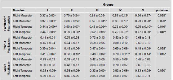
*Statistically significant difference by ANOVA (p < 0.05)
**Different letters indicate statistically significant difference according to Duncan's test (p< 0.05).
During the clinical condition of habitual and nonhabitual mastication, group I (children) had lower electromyographic activity in all analyzed muscles. There were changes in the performance of masseter and temporalis muscles, bilaterally in the group IV (adults) during the conditions of habitual and non-habitual mastication. It was observed that there was an increase in masticatory function in GII (teenagers) compared to GI (children) and a decrease in function in GIII (young adults) for all the muscles analyzed. GV (elderly) had increased electromyographic activity during the non-habitual mastication function for the masseter muscles, bilaterally and in mastication (peanuts) to the right and left temporal muscles, with statistically significant difference.
The results of the Multivariate Analysis (General Linear Models) are shown in Table 2. Group effects were observed for the mastication of parafilm MR (non-habitual) and of raisins (habitual), but there was no interaction between group and gender for any kind of mastication.
Table 2: F values for the General Linear Models across Group and Gender.

DISCUSSION
The human body undergoes continuous transformations in its form and function as from birth. Over time, there is a decrease in the ability of the physiological system to perform its functions, which exacerbates the effects of ageing on the body structures15. A careful review of the standards related to jaw dynamics, muscle function, masticatory function and occlusion may provide valuable information for a better understanding of the diseases that affect the stomatognathic system throughout life.
Despite the limitations of electromyography that have not yet been solved, such as skin tissue conductibility16, position of the electrodes and the concurrent activities of nearby muscles17, protocol and procedures for electrode placement, electrode shape, size and location, surface electromyography has provided the means to further our understanding of the physiology of the masticatory system5. The maintenance of healthy teeth and muscles is extremely important for mastication as well as in promoting a healthy functional state of the stomatognathic system2,18.
In the present study, the evaluation of masticatory process by means of electromyography was the method of choice. It is effective for the diagnosis, evaluation and monitoring of the alterations which, over time, affect the muscle performance during mastication. It is also important for the investigation of functional integrity and treatments; however, it will not demonstrate the final product of mastication19-22. Several artificial and natural foods are used for the evaluation of the masticatory function21. In our study, the parafilm was used as simulator food in the chewing test, and peanuts (hard food) and raisins (soft food) were selected as natural foods. The natural foods used in masticatory performance tests are consumed on a daily basis, and people are accustomed to their consistency, in contrastto artificial foods23. Individuals consume food with a wide variety of consistencies and hardness, therefore, any method used for the evaluation of the masticatory function with certain food, regardless of whether it is natural or artificial, can only measure its mastication potential.
The type and consistency of food, neuromuscular quality, age, and number of teeth influence the duration and frequency of the masticatory process24. During mastication, the muscles involved spend energy on grinding the food and then swallowing20. Studies have shown that age and gender are important factors that influence the resistance of jaw lifting muscles in terms of the masticatory process18. The results of this study confirmed that children in GI (7 to 12 years) had less efficient electromyographic activity, both in the habitual and non-habitual mastication, compared to the other age groups. This could be due to the muscular development in children, which generates less muscle strength and limited mandibular movements compatible with their age, showing that the masticatory pattern alters during growth, probably as a result of anatomical changes and the development of the nervous system25,26. The oral structures acquire morphological and functional characteristics different from those of younger individuals27.
Many studies show that there is a significant decrease in muscle mass and performance in the elderly, while the stomatognathic system, which is independent, can undergo alterations with an increase in the number of masticatory cycles for the food to be ground; however the system has the ability to adapt, and the masticatory function can be maintained or even increased with age, as a result of the recruitment of muscle fibers to maintain the stability of masticatory function28.
Elderly individuals without health problems, with all natural teeth, habitual occlusion and not taking any drugs that modify the electromyographic activity of the masseter and temporalis muscles may show increased masticatory process29. These data are consistent with those of the present study where Group V (elderly people), when compared to Group I (children), Group II (teenagers), Group III (young adults) and Group IV (adults) showed increased masticatory efficiency.
Most of the time during this study, the results showed less activation of the temporalis muscles during mastication of soft food (raisins) in all groups. This would not be expected with soft food, since the temporalis muscles are functionally more active during fast mandibular movements; however, with regard to hard food (peanuts), which requires chewing force, the masseter muscles should be more active1, according to the results found in the present study.
With the enrollment of a healthy, heterogeneous population aged between 7 and 80 years, our study provides data that may serve as standards for the Brazilian population in terms of the efficiency of the masticatory cycle and the interaction with the group factor. The understanding and evaluation of the efficiency of the masticatory cycle using the electromyographic activity are of great importance to dentistry, since they provide the diagnosis, evaluation and monitoring of alterations that may affect the performance of the muscles during mastication over the years, and are also essential for the investigation of the oral health condition and the treatments proposed1, 29.
CONCLUSION
With the enrollment of a healthy population aged between 7 and 80 years, our study provides data that may serve as standards for the Brazilian population in terms of the masticatory proces. The results of this study indicate that age and gender are associated with structural and functional alterations in the masticatory muscles.
ACKNOWLEDGEMENTS
This study was supported by a grant from the ‘‘Fundacao de Amparo a Pesquisa - FAPESP''
1. Siessere S, de Albuquerque Lima N, Semprini M, de Sousa LG, Paulo Mardegan Issa J, Aparecida Caldeira Monteiro S, Cecilio Hallak Regalo S. Masticatory process in individuals with maxillary and mandibular osteoporosis: electromyographic analysis. Osteoporos Int 2009;20:1847-1851. [ Links ]
2. Pereira LJ, Duarte Gaviao MB, Van Der Bilt A. Influence of oral characteristics and food products on masticatory function. Acta Odontol Scand 2006;64:193-201. [ Links ]
3. Galo R, Vitti M, Santos CM, Hallak JE, Regalo SC. The effect of age on the function of the masticatory system-an electromyographical analysis. Gerodontology 2006;23:177-182. [ Links ]
4. Regalo SC, Vitti M, Semprini M, Rosa LB, Martinez FH, Santos CM, Hallak JE . Electromyographic analysis of the masseter and temporal muscles in oralized deaf individuals. Electromyogr Clin Neurophysiol 2006;46:217-222. [ Links ]
5. Castroflorio T, Bracco P, Farina D. Surface electromyography in the assessment of jaw elevator muscles. J Oral Rehabil 2008;35:638-645. [ Links ]
6. Piancino MG, Isola G, Merlo A, Dalessandri D, Debernardi C, Bracco P. Chewing pattern and muscular activation in open bite patients. J Electromyogr Kinesiol 2012;22:273-279. [ Links ]
7. Andrade AS, Gaviao MB, Derossi M, Gameiro GH. Electromyographic activity and thickness of masticatory muscles in children with unilateral posterior crossbite. Clin Anat 2009;22:200-206. [ Links ]
8. Cecilio FA, Regalo SC, Palinkas M, Issa JP, Siessere S, Hallak JE, Machado-de-Sousa JP, Semprini M. Ageing and surface EMG activity patterns of masticatory muscles. J Oral Rehabil 2010;37:248-255. [ Links ]
9. Kanayama H, Masuda Y, Adachi T, Arai Y, Kato T, Morimoto T. Alteration of masticatory muscle EMG activities during chewing after a reversible bite-raising in guinea pigs. Arch Oral Biol 2011;56:793-798. [ Links ]
10. Paphangkorakit J, Chaiyapanya N, Sriladlao P, Pimsupa S. Determination of chewing efficiency using muscle work. Arch Oral Biol 2008;53:533-537. [ Links ]
11. Andrade Ada S, Gaviao MB, Gameiro GH, De Rossi M. Characteristics of masticatory muscles in children with unilateral posterior crossbite. Braz Oral Res 2010;24:204-210. [ Links ]
12. Finni T, Cheng S.Variability in lateral positioning of surface EMG electrodes. J Appl Biomech 2009;25:396-400. [ Links ]
13. Hermens HJ, Freriks B, Disselhorst-Klug C, Rau G. Development of recommendations for SEMG sensors and sensor placement procedures. J Electromyogr Kinesiol 2000; 10:361-374. [ Links ]
14. Borges Tde F, Regalo SC, Taba M Jr, Siessere S, Mestriner W Jr, Semprini M. Changes in masticatory performance and quality of life in individuals with chronic periodontitis. J Periodontol 2013;84:325-331. [ Links ]
15. Galo R, Vitti M, Mattos Mda G, Regalo SC. Masticatory muscular activation in elderly individuals during chewing. Gerodontology 2007;24:244-248. [ Links ]
16. Fujita T, Fujii Y, Okada SF, Miyauchi A, Takagi Y. Fall of skin impedance and bone and joint pain. J Bone Miner Metab 2001;19:175-179. [ Links ]
17. Lund JP, Widmer CG. Evaluation of the use of surface electromyography in the diagnosis, documentation, and treatment of dental patients. J Craniomandib Disord 1989; 3:125-137. [ Links ]
18. Hatch JP, Shinkai RS, Sakai S, Rugh JD, Paunovich ED. Determinants of masticatory performance in dentate adults. Arch Oral Biol 2001;46:641-648. [ Links ]
19. Shiau YY, Peng CC, Wen SC, Lin LD, Wang JS, Lou KL. The effects of masseter muscle pain on biting performance. J Oral Rehabil 2003;30:978-984. [ Links ]
20. Feine JS, Lund JP. Measuring chewing ability in randomized controlled trials with edentulous populations wearing implant prostheses. J Oral Rehabil 2006;33:301-308. [ Links ]
21. Harridge SD, White MJ, Carrington CA, Goodman M, Cummins P. Electrically evoked torque-velocity characteristics and isomyosin composition of the triceps surae in young and elderly men. Acta Physiol Scand 1995;154:469-477. [ Links ]
22. Fontijn-Tekamp FA, van der Bilt A, Abbink JH, Bosman F. Swallowing threshold and masticatory performance in dentate adults. Physiol Behav 2004; 83: 431-436. [ Links ]
23. Santos CM, Vitti M, Matsumoto W, Berro RJ, Semprini M, Hallak JEC, et al. Using overdenture on implants and complete denture: effects on postural maintenance of masticatory musculature. Braz J Oral Sci 2007;7:1550-1504. [ Links ]
24. Akeel R, Nilner M, Nilner K. Masticatory efficiency in individuals with natural dentition. Swed Dent J 1992; 16:191-198. [ Links ]
25. Papargyriou G, Kjellberg H, Kiliaridis S. Changes in masticatory mandibular movements in growing individuals: a six-year follow-up. Acta Odontol Scand 2000; 58: 129-134. [ Links ]
26. Palinkas M, Nassar MS, Cecilio FA, Siessere S, Semprini M, Machado-de-Sousa JP, Hallak JE, Regalo SC. Age and gender influence on maximal bite force and masticatory muscles thickness. Arch Oral Biol 2010;55:797-802. [ Links ]
27. Peyron MA, Blanc O, Lund JP, Woda A. Influence of age on adaptability of human mastication. J Neurophysiol 2004;92:773-779. [ Links ]
28. Alajbeg IZ, Valentic-Peruzovic M, Alajbeg I, Cifrek M. The influence of age and dental status on elevator and depressor muscle activity. J Oral Rehabil 2006;33:94-101. [ Links ]
29. Mishellany-Dutour A, Renaud J, Peyron MA, Rimek F, Woda A. Is the goal of mastication reached in young dentates, aged dentates and aged denture wearers? Br J Nutr 2008;99:121-128. [ Links ]

