


Servicios Personalizados
Revista

Articulo
 Inglés (pdf)
 Articulo en XML
 Referencias del artículo
 Enviar articulo por email
Indicadores
-
 Citado por SciELO
Links relacionados
-
 Similares en
SciELO
Similares en
SciELO
Compartir

 Permalink
Acta Odontológica Latinoamericana
versión On-line ISSN 1852-4834
Acta odontol. latinoam. vol.27 no.3 Buenos Aires dic. 2014
ARTÍCULOS ORIGINALES
Morphological changes related to age in mesial root canals of permanent mandibular first molars
Omar A Gani1, Claudio F Boiero1, Carolina Correa1, Ivana Masin1, Ricardo Machado2, Emmanuel JNL Silva3, Luiz Pascoal Vansan2
1 Departamento de Endodoncia, Córdoba National University (UNC), Córdoba, Argentina
2 Departamento de Endodontia, São Paulo University (FORP/USP), Ribeirão Preto, Brazil
3 Health Science Center, Grande Rio University (UNIGRANRIO), Rio de Janeiro, Brazil
CORRESPONDENCE Dr. Emmanuel JNL da Silva Rua Herotides de Oliveira 61/902 Icarai, Niteroi, RJ, Brazil nogueiraemmanuel@hotmail.com
ABSTRACT
The aim of this study was to evaluate age-related morphological canal changes in mesial root canals of mandibular first molars of known ages. Fifty-six specimens were selected for this study and distributed into the following four age groups (n. 14): a) Group of children under 13 years, b) Group of adolescents (from 14 to 19 years), c) Group of young adults (from 20 to 39 years) and d) Group of older adults (over 40 years). The specimens were in perfect condition because after extraction they were carefully cleaned, sterilized, identified and stored in water. In order to improve the cleaning, they were placed in 1% sodium hypochlorite solution for four hours and rinsed in 10 vol. hydrogen peroxide for 8 hours. After that, a clearing technique was performed to illustrate root canal anatomy. Digitalized images of all samples were obtained by use of a stereomicroscope. Canals were noticeably simpler in older adults: they were sharply defined and narrow, sometimes too narrow. Calcification nuclei were not found and there were only a few remains of internuclear spaces. The canal system appeared cleaner, clearer and more sharply defined than in the other age groups. It may be concluded that there is a correlation between aging and morphological changes in the mesial root canals of mandibular first molars.
Keywords: Anatomy; Molar; Morphology.
RESUMO
Alterações morfológicas relacionadas à idade em canais radiculares mesial de primeiros molares inferiores permanentes
O objetivo do presente estudo foi avaliar as alteracoes morfologicas relacionadas com a idade em canais radiculares mesial de primeiros molares inferiores. Cinquenta e seis especimes foram selecionados para este estudo. Os especimes foram distribuidos em quatro grupos etarios (n. 14): a) Grupo de criancas menores de 13 anos, b) grupo de adolescentes (de 14 a 19 anos), c) Grupo de jovens adultos (de 20 a 39 anos ) e d) Grupo de adultos (acima de 40 anos). Apos as extracoes os elementos foram cuidadosamente limpos, esterilizados, identificadas e armazenadas em agua. A fim de melhorar a limpeza, foram colocados numa solucao de hipoclorito de sodio a 1%, durante quatro horas e enxaguados em 10 vol. peroxido de hidrogenio durante 8 horas. Depois, uma tecnica de diafanizacao foi realizada para ilustrar a anatomia do canal radicular. As imagens digitalizadas de todas as amostras foram obtidas atraves da utilizacao de um estereoscopio. Os canais foram visivelmente mais simples em adultos mais velhos: eles foram bem definidas e estreito, por vezes, demasiado estreito. Nucleo de calcificacao nao foi encontrado e havia apenas alguns restos de espacos internucleares. O sistema de canal apareceu mais limpa, mais clara e mais bem definida do que nas outras faixas etarias. Pode-se concluir que ha uma correlacao entre as alteracoes do envelhecimento e morfologicas nos canais radiculares mesial de primeiros molares inferiores.
Palavras-chave: Anatomia; Molar; Morfología.
INTRODUCTION
The pulp dentinal complex is capable of responding to a variety of stimuli over time. These stimuli can be physiological -relating to the normal stresses to which a tooth would be exposed over a lifetime-, or pathological -due to caries, tooth surface loss, or restorative treatment1. One of the most obvious features of aging is a reduction in size of the pulp chamber caused by the continual secretion of dentinal matrix (physiological secondary dentinogenesis) by odontoblasts2. This process tends to narrow the originally wide-open root apex; with aging, the apical deposition of secondary dentin and cementum increases, and circulation and innervation are compromised. Teeth reflect the biological or physiological age of the individual, and variations caused by genetic factors and chewing habits can influence tooth anatomy3.
The complexity of root canal systems and internal morphology has been directly correlated with endodontic treatment planning, therapy and outcome. Mandibular first molars are the first permanent teeth to erupt, often requiring endodontic care due to early caries4. Typically, mandibular molars have two well-defined roots: a mesial root characterized by a flattened mesiodistal surface and widened buccolingual surface, and a distal root mostly straight with a wide oval canal or two round canals5. However, many variations exist regarding its root and root canal anatomy, thus necessitating critical evaluation of each individual case for variations6-8.
In this study, we evaluated age-related morphological canal changes in mesial root canals of mandibular first molars of known ages. The tested null hypothesis was that there is no difference in the anatomy of mesial root canals of mandibular first molars at different ages.
MATERIALS AND METHODS
The Ethics Committee approved this study. Fiftysix first mandibular molars were provided by the Tooth Bank of Cordoba National University. The teeth were selected for this study considering age, integrity of mesial root and complete root formation. In order to improve cleaning, teeth were placed in 1% sodium hypochlorite solution for four hours and rinsed in 10% hydrogen peroxide for 8 hours. The specimens were distributed into the following four age groups (n=14): a) Group of children under 13 years, b) Group of adolescents (from 14 to 19 years), c) Group of young adults (from 20 to 39 years) and d) Group of older adults (over 40 years). After coronal access, mesial roots were placed in a plastic tube connected to a vacuum and the tuberoot junction was sealed with wax. Then, a luer-lock syringe with a 27-gauge endodontic needle was used to inject Indian ink into the mesial canals while vacuum was applied. This operation was repeated until ink emerged from the foramen. After drying, the teeth were decalcified in 5% nitric acid for 28 to 30 hours. After thorough washing of the decalcified teeth in running tap water for 4 hours, the samples were dehydrated in ascending concentrations of ethanol (70%, 80%, 95% and 100%) for 1 day, and the samples were rendered transparent by immersion in methyl salicylate for 2 days.
Digitalized images of all samples in 5 different angles were obtained by use of a stereomicroscope (Olympus Co, Tokyo, Japan) under 5X magnification in order to analyze the anatomy of each specimen of the four groups.
RESULTS
Group of children under 13 years
The images of the canal system in children were highly variable. Sometimes there were single, large, triangular-shaped canals with the vertex ending in a single apical foramen (Fig. 1A, B) while other times the entire canal, including the apical section, was ribbon- shaped (Fig. 1C, D). The canals often had blister- shaped dilatations, particularly in the coronal and middle sections of the roots (Figs. 1E, F, G, H, I, J). There were often unstained circular or oval-shaped areas within the canal, which were sometimes very small (Fig. 1E) and sometimes large (Fig. 1F, G, H). We call them "calcification nuclei". In most cases the calcification nucleus was single and located at the widest part of the canal, which would later allow its surface to increase, tending clearly to determining two canals. Thus, if the shape of canal tended to be triangular (Fig. 1A), the calcification nucleus developed in its coronal third (Fig. 1F, G), and if the canal was somewhat dilated in the middle third (Fig. 1B), the calcification nucleus began there and then extended in all directions, particularly towards the apex, foreshadowing the presence of two canals joining up to form a single foramen (Fig. 1H).

Fig. 1: Representative images from children under 13 years.
Ribbon-shaped canals had quite particular anatomical features. The bifurcations and trifurcations at the apical third of the canal and even the presence of veritable apical deltas seem to be very frequent (Figs. 1C, D, I, K), more so than in triangular canals. Moreover, it should be highlighted that the calcification nucleus in these ribbon-shaped canals began at the apical third, foreshadowing the presence of the two canals ending in different foramina (Fig. 1I, K).
Group of adolescents (14 to 19 years)
The anatomical appearance of canals in adolescents was similar to that in children. The only difference between a triangular canal in adolescents (Fig. 2A) and children was that the former had a larger calcification nucleus and more clearly defined anatomy. The presence of two, three or more calcification nuclei was fairly frequent in adolescents, whether the canals ended in one or two foramina. Nuclei were predominantly oval-shaped, and their sizes varied noticeably. Their distribution in the canal depended on their number. There were linear spaces between them, which we call internuclear spaces (Fig, 2B, C), of widely varying appearance: they could be wide or narrow; horizontal, oblique or vertical; one, two or several, according to the number of nuclei.
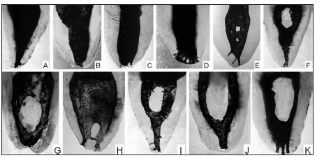
Fig. 2: Representative images from adolescents (14 to 19 years).
The development of a calcification nucleus at the apical section of the canal often led to bifurcation of the canal, ending in two individual foramina, which could either be very close to each other (Fig. 2C) or very far apart from each other (Fig. 2D, E, F, G). In the latter case, the apical bifurcations were very evident and sometimes the calcification nucleus between them was formed by the fusion of two or more smaller nuclei, with only few remains of the spaces. In contrast, calcification nuclei were not found in the middle and coronal canal sections of the canals.
Group of young adults (20 to 39 years)
In young adults, the canals were more clearly defined than in adolescents. Nevertheless, there were still calcification nuclei and spaces separating them, often forming a complex system (Fig. 3A, B). Vertical internuclear spaces joining up with other spaces or canals after a short length were outstanding due to their clear definition.

Fig. 3: Representative images from young adults (20 to 39 years).
Group of older adults (over 40 years)
Canals were noticeably simpler in older adults: they were sharply defined and narrow, sometimes too narrow. Calcification nuclei were not found and there were only a few remains of internuclear spaces. The canal system appeared cleaner, clearer and more sharply defined than in the other age groups (Fig. 4A, B, C).

Fig. 4: Representative images from older adults (over 40 years).
DISCUSSION
Knowledge of the influence of age on root canal anatomy is needed to improve the cleaning and shaping process in endodontic treatment of patients of different ages. This study investigated age-related changes in mesial root canals of mandibular first molars. The results of this study showed a reduction of the canal, and several other differences, by comparing the canal anatomy of teeth of different ages. Therefore, the null hypothesis was rejected. Previous studies correlating patient age with root canal anatomical changes1-3,9,10 have shown similar results, demonstrating that the canal undergoes reduction over the years. In children, there were cases in which the canal was single throughout its length, wide at the coronal section, narrowing gradually towards the apex and ending in a single foramen. In other cases, the width and ribbon shape were even along the entire length of the canal, even in the apical section. The results in this group are probably related to the dentin deposition process, which, over time, reduces the main canal lumen and significantly changes the shape of the root canal, including a partial separation with the presence of an isthmus10. Isthmuses cause great difficulty in the cleaning and shaping process, as they have been shown to be inaccessible to conventional hand and rotary instrumentation11,12.
Similar anatomy was observed in adolescents, although the size and number of calcification nuclei was greater. In certain cases, the internuclear spaces formed a real systems or plexus whose complexity depended on the number of calcification nuclei. In this group, there were frequently calcification nuclei dividing the canals in the apical section, some of which seemed to be formed by the fusion of two or more nuclei. On the other hand, nuclei in the middle and coronal sections of the canal were noticeably absent. Nevertheless, to compensate, it would appear that the apical calcification nucleus develops upward, increasing the division between the canals. The ending of both canals in independent foramina contributes to an interesting anatomical feature, because despite the lack of endodontic maturation still evident in adolescence, the apical portion of the canals showed some degree of anatomical definition and above all, a narrowing of both canals enabling better quality in instrumentation and canal filling aspects, which have been reported previously13. These observations allow us to state that in the mesial root of mandibular molars, a certain degree of endodontic maturation can only be spoken of in late adolescence, and not in all cases, because sometimes the process continues into the young adult stage. In young adults, there is no doubt that the signs of endodontic maturation are clearer. Nevertheless, there are still calcification nuclei and spaces separating them, although these spaces are smaller and sometimes fewer. Figun & Garino14, referring to them as inter-canal communications and disregarding age, report that there may be 4 or 5 in a single canal and their sizes may range in diameter from filiform to 1mm. They describe their shape and direction as straight, arched or italic-S-shaped, and according to the communicating canals, classify them as primary, secondary or tertiary. In contrast, Peiris et al.15 reported, without providing details, that the prevalence of inter-canal communications is low at young and old ages and high at intermediate ages, which, in very general terms, is what our study found. Vertical internuclear spaces are particularly outstanding in young adults, and have not been reported by the abovementioned authors, although they have sometimes been considered as additional canals16, when in fact they simply show immaturity, since they will soon be vestigial or will have disappeared entirely.
In older adults, we did not find any calcification nuclei or internuclear spaces, although there were a few remains. The anatomical features usually found in canals of older adults are those which could already be foreseen in children, began to take shape in adolescents and particularly in young adults, to reach full maturity in older adults. These observations are similar to those in a previous study15, which reported that in lower molars, the canals are large up to the age of 11 to 15 years, and the internal shape is defined between 30 and 40 years. The success of endodontic treatment in adults, particularly in older adults, might be due to the fact that the pulp cavity becomes narrower with age, enabling better shaping and filling procedures17. Therefore, successful endodontics can be achieved in older adults with special attention to diagnosis, good quality radiographs and an adequate technique to overcome the challenges posed by anatomy changes of the root canal system.
Under the present experimental framework and with the limited sample size, a correlation between aging and morphological changes was observed in the mesial root canals of mandibular first molars.
1. Burke FM, Samarawickrama DY. Progressive changes in the pulpo-dentinal complex and their clinical consequences. Gerodontology 1995;12:57-66. [ Links ]
2. Morse DR, Esposito JV, Schoor RS. A radiographic study of aging changes of the dental pulp and dentin in normal teeth. Quintessence Int 1993;24:329-333. [ Links ]
3. Lopez Nicolas M, Morales A, Luna A. Morphometric study of teeth in age calculation. J Forensic Odontostomatol 1993;11:1-8. [ Links ]
4. Berkovitz BK, Holland GR, Muxham BJ. Tooth morphology. In: Berkovitz BK, Holland GR, Muxham BJ, editors. Oral anatomy Histology and Embryology. London: Wolf; 1992. p. 24-43. [ Links ]
5. Skidmore AE, Bjorndal AM. Root canal morphology of the human mandibular first molar. Oral Surg Oral Med Oral Pathol 1971;32:778-784. [ Links ]
6. Chandra SS, Chandra S, Shankar P, Indira R. Prevalence of radix entomolaris in mandibular permanent first molars: a study in a South Indian population. Oral Surg Oral Med Oral Pathol Oral Radiol Endod 2011;112:e77-82. [ Links ]
7. Kim SY, Yang SE. Cone-beam computed tomography study of incidence of distolingual root and distance from distolingual canal to buccal cortical bone of mandibular first molars in a Korean population. J Endod 2012;38:301-304. [ Links ]
8. Silva EJ, Nejain Y, Silva AI, Haiter-Neto F, Cohenca N. Evaluation of root canal configuration of mandibular molars in a Brazilian population using cone-beam computed tomography. an in vivo study. J Endod 2013;39:849-852. [ Links ]
9. Hess W, Zurcher E, The anatomy of the root canals of the teeth of the permanent dentition and the anatomy of the root canals of the deciduous dentition and the first permanent molars, Basle, Sons and Danielson, London, 1925. [ Links ]
10. Artal N, Gani O. Endodontic Anatomy of the root canals of lower incisors. Acta Odontol Latinoam 2000;13:39-49. [ Links ]
11. Adcock JM, Sidow SJ, Looney SW, McNally K, Lindsey K, Tay FR. Histologic evaluation of canal and isthmus debridement efficacies of two different irrigant delivery techniques in a closed system. J Endod 2011;37:544-548. [ Links ]
12. Versumer J, Hulsmann M, Schafers F. A comparative study of root canal preparation using Profile .04 and Lightspeed rotary Ni-Ti instruments. Int Endod J 2002;35:37-46. [ Links ]
13. Gani O, Visvisian C, Rodrigo R, David O. Anatomia radiografica de los conductos radiculares del primer molar inferior con especial referencia a sus curvaturas. Endodoncia 1993;11:64-73. [ Links ]
14. Figun ME, Garino RR. Anatomia Odontologica. Buenos Aires, El Ateneo 2006 pag. 399-518. [ Links ]
15. Peiris R, Takahashi M, Sasaki K, Kanazawa E. Root and canal morphology of permanent mandibular molars in a Sri Lankan population. Odontology 2007;95:16-23. [ Links ]
16. Pomeranz HH, Edelman DL, Goldberg MG. Treatment considerations of middle mesial canal of mandibular first and second molars. J Endod 1981;7565-7568. [ Links ]
17. Allen PF, Whitworth JM. Endodontic considerations in the elderly. Gerodontology 2004;21:185-194. [ Links ]

