


Servicios Personalizados
Revista

Articulo
 Inglés (pdf)
 Articulo en XML
 Referencias del artículo
 Enviar articulo por email
Indicadores
-
 Citado por SciELO
Links relacionados
-
 Similares en
SciELO
Similares en
SciELO
Compartir

 Permalink
Acta Odontológica Latinoamericana
versión On-line ISSN 1852-4834
Acta odontol. latinoam. vol.29 no.2 Buenos Aires set. 2016
ARTÍCULOS ORIGINALES
Characterization of third molar morphometric variables
Pablo W. Trinks1, María Belén Grifo1, Fernando Pari1, Mariano A.R. Amer1, Gabriel A. Sánchez2
1 Department of Anatomy, School of Dentistry, University of Buenos Aires, Argentina
2 Department of Biophysics, School of Dentistry, University of Buenos Aires, Argentina.
CORRESPONDENCE Prof. Dr. Mariano A. R. Amer Catedra de Anatomia. Facultad de Odontologia UBA. M T de Alvear 2142, 1st Floor B, Buenos Aires, 1122AAH, Argentina marianoamer@gmail.com
ABSTRACT
The third molar is a tooth of anatomical, surgical, prosthetic and forensic dental interest. However, there is currently a lack of updated data regarding its morphology. The aim of this study was to determine the morphometric features of third molars and their predictive capability as regards dental arch and side. Two calibrated operators (ƙ = 0.83) determined the cervicalocclusalvestibular (COV), cervicalocclusalpalatal (COP) and occlusalapical (OA) distances, mesiodistal (MD), and vestibularpalatal (VP) diameters, number of roots (R) and number of cusps (C) of 961 cadaveric third molars, both upper (n = 462) and lower (n = 499), using a CONCOR 050 thin mandible caliper (resolution 0.01 mm). Median and range for each variable were calculated and compared using Mann Whitney nonparametric test (p < 0.05). Multivariate cluster analysis was used to determine the predictive capability of each variable for dental arch and side. For upper molars (UM), 50.6% were from the right side (RS) and 49.4% from the left side (LS), while for lower molars (LM), 60.9% were from the RS and 39.1% from the LS. No significant difference was found in the study variables in LM according to side. For UM, MD diameter (10.90 mm), COP(7.42 mm) distance and number of R (3) were significantly higher (p < 0.05) forRS, and number of C (3) was higher (p < 0.0001) for LS. They were also significant predictive grouping factors for side. For dental arch, OA (17.84 mm) and COV (7.60 mm) distances, MD (11.26 mm) diameter and the number of C (5) were significantly higher (p < 0.0001) for LM, while VP (10.84 mm) and COP (7.34 mm) distances, and the number of R (3) were significantly higher (p < 0.0001) for UM. These variables were significant predictive factors for dental arch. Despite the morphometric heterogeneity of third molars, there are intrinsic parameters with predictive capability for dental arch and side, but it would be advisable to supplement this study with data from topographic occlusal variables in order to validate their predictive capability.
Key words: Tooth; Morphology; Third molar.
RESUMEN
Caracterización de variables morfométricas del tercer molar
El tercer molar es una pieza dentaria de interés odontológico anatómico, quirúrgico, endodóntico, protético y forense. Sin embargo, no hay disponibles hoy en día datos morfométricos actualizados del molar. El objetivo del trabajo fue determinar las características morfométricas de terceros molares y establecer el carácter predictivo de las mismas en cuanto a arco dental y lado. Dos operadores calibrados (ƙ = 0.83) determinaron la longitud oclusocervical vestibular (OCV) y palatina (OCP), oclusoapical (OA), diámetro mesiodistal (MD), diámetro vestíbulopalatino (VP), número de raíces (R) y número de cúspides (C), de 961 terceros molares cadavéricos, superiores (n = 462) e inferiores (n = 499), mediante el uso de un calibre de mandíbula fina CONCOR 050 (resolución 0.01 mm). Calculamos mediana y rango para cada variable y las comparamos haciendo uso de la prueba de Mann Whitney (p < 0.05). Utilizamos el análisis de cluster para determinar el valor predictivo de cada variable en cuanto a arco y lado. De los molares superiores (MS), 50.6% correspondió al lado derecho (LD) y 49.4% al lado izquierdo (LI). De los inferiores (MI), 60.9% correspondió al LD y 39.1% al LI. No hallamos diferencias significativas para las variables en estudio para los MI según su lado. Para los MS, el MD (10.90 mm), la OCP (7.42 mm) y el R (3) resultaron significativamente mayores (p < 0.05) para el LD, y el C (3), mayor (p < 0.0001) para el LI; y, además evidenciaron significancia como factores predictivos de agrupamiento para la predicción del lado. En relación al arco, la OA (17.84 mm), la OCV (7.60 mm), el MD (11.26 mm) y el C (5), resultaron significativamente mayores (p < 0.0001) en los MI, mientras que el VP (10.84 mm), la OCP (7.34 mm) y el R (3) fueron significativamente mayores (p < 0.0001) en MS. Dichas variables evidenciaron significan cia como factores predictivos para el arco. Pese a la heteroge neidad morfométrica del tercer molar, existen parámetros característicos con valor predictivo para el arco y lado, aunque sería recomendable complementar el estudio con variables topográficas oclusales a fin de validar la capacidad predictiva de los mismos.
Palabras clave: Diente; Morfología; Tercer molar.
INTRODUCTION
The different types of teeth perform different functions, such as mastication, speech, esthetics and preservation (maintenance of the integrity of periodontal tissues). Teeth are important in preserving the balance and harmony of the dental arch and the dental and masticatory systems1. The third molar fulfills all the abovementioned functions; however, it has specific features. Its calcification begins at 9 years of age, and it usually erupts between 18 and 25 years of age. After its eruption, the calcification and conformation of the third molar is completed over a period of 1 to 1.5 years2. Given the omnivorous diet of humans, like the rest of the molars, it performs cutting, friction and grinding functions during mastication3. The third molar shows more morphological variations than any other tooth4-6. The occlusal surface is rough due to the presence of short, shallow grooves; roots are usually short, fused and curved distally at the apical third7.
Ontogenic and phylogenetic studies suggest that, in modern man, the molar series is decreasing in size distally as a consequence of a reduction in the masticatory function, with a decrease in the number and size of teeth, number of molar cusps and size of jaws, over the ages8. However, the opposite is observed in monkeys and primitive races, the consequence of which is that the third molar is smaller than the first and second molars and is usually retained or absent9. In dentistry, not only is the third molar a tooth of anatomical interest, but also of surgical, endodontic, prosthetic and forensic interest. The morphometric study of this particular tooth is of vital importance for said specialties. The study of the retained third molar, indication for its removal and potential complications associated with it require precise morphometric assessment of the tooth in question10. This is emphasized by the fact that some morpho metric characteristics of the lower third molar have been associated with a higher incidence of mandibular angle and condylar fractures11. The morphometric characteristics of this tooth have also been recently included as a measure of interest in predictive models regarding the duration of third molar surgery12. Moreover, the morphometric study of this particular tooth is of interest in the determination and validation of parameters concerning the measurements and architecture of the dental arches13. In the field of forensic dentistry, the morphometric study of the third molar is vital in dental recognition of fossil remains14. From the prosthetic point of view, the need for preservation of this tooth is not infrequent, prior to which the relevant endodontic therapy should be performed, which requires accurate knowledge of the tooth and root morphology15.
Despite the importance of studying dental mor phometry and topography14, 16, there is no consistent update of morphometric data of the third molar. The available national literature only refers to data published almost four decades ago17. The advent of new techniques for morphometric and topographical study of teeth7,16provides favorable prospects for updating such data. The incorporation of mathematical tools for creating computational models predictive of the occlusal morphology and topography of teeth and the jaw bone constitutes a broad field where updated morphometric data for the third molar are to be specifically applied18. The pressing need for updating the morphometric data of the third molar and its subsequent statistical analysis is the main reason for undertaking this research, and the aim of this study was to identify the morphometric characteristics of the upper and lower third molar and their predictive capability as regards dental arch and side.
MATERIALS AND METHODS
This research was conducted as a descriptive study. Nevertheless, analytic comparison and predictive capability of the observed dental parameters have been included herein. The study population consisted of cadaveric teeth, including bilateral upper and lower third molars from the ossuary of the Department of Anatomy of the School of Dentistry, University of Buenos Aires, Argentina. The molars were prepared for handling according to the requirements set by the Biosafety Commission of the School of Dentistry. Nine hundred and sixtyone third molars (462 upper and 499 lower) were studied. Molars with abnormal crown or roots, or any that were partially destroyed or filled were not included in the study population. The morphometric determinations comprised the occlusalapical (OA), cervicalocclusalvestibular (COV) and cervicalocclusalpalatal (COP) lengths (Fig. 1), and mesiodistal (MD) and vestibularpalatal (VP) diameters (Fig. 2). Measurement data corresponding to the different tooth morphometric parameters were determined in mm by means of a CONCOR 050 mm thin mandible digital caliper (resolution: 0.01 mm) and a TESA 25/50 (025 mm) digital micrometer. The measurement data corres ponding to the number of roots (R) and cusps (C), of the occlusal surface of the third molars were determined macroscopically by dental recognition. Data were collected by two calibrated operators (Cohens kappa coefficient of 0.83).
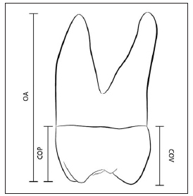
Fig. 1: Proximal view of a generic third molar. The drawing illustrates the occlusalapical (OA), cervicalocclusalvestibular (COV) and cervical occlusal palatal (COP) lengths.
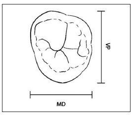
Fig. 2: Occlusal view of a generic third molar.The drawing illustrates the mesiodistal (MD) and vestibularpalatal (VP) diameters.
The descriptive statistical analysis included the calculation of median and range for each studied variable. Differences in the morphometric parameters were tested for significance by Mann Whitney test using GRAPHPAD Prism version 5.03 for Windows (GraphPadSoftware, San Diego, CA, USA). Multivariate cluster analysis of the abundance levels of dental measurements was used to determine the hierarchical dissimilarity and predictive capability of each variable as for dental arch and side. The level of significance used was p < 0.05.
RESULTS
For upper molars, 50.6% were from the right side and 49.4% from the left side, while for lower molars, 60.9% were from the right side and 39.1% from the left side.
The boxplot in Fig. 3 shows that the upper third molars have a statistically significantly higher number of cusps (Mann Whitney U = 22230, p = 0.0003) on the left side (median, range: 3, 36) than on the right side (3, 35), whereas the number of roots was statistically significantly higher (Mann Whitney U = 24650, p = 0.028) in molars on the right side (3, 15) than on the left (3, 14). The OA length (Fig. 4 A) and the VP and COV distances did not differ significantly between sides (Fig. 4 B); however, the median of the MD and COP measurements of right molars were significantly higher (Mann Whitney U = 23540, p = 0.0287 for MD measurements and Mann Whitney U = 22820, p = 0.0089 for COP measurements, respectively, than those of left molars (Fig. 4 B). For right molars, measurements (median, range) were: OA:17.45, 11.1222.56 mm; VP: 10.90, 8.8113.27 mm; MD: 9.37, 7.2911.27 mm; COV: 7.15, 3.558.77 mm; COP: 7.42, 4.299.79 mm. For left molars, they were: OA: 17.35, 11.4524.00 mm; VP: 10.80, 8.1613.68 mm; MD: 9.16, 6.5211.18 mm; COV: 7.06, 3.878.81 mm; COP: 7.26, 4.289.03 mm. The lower left and right third molars showed no significant differences in the number of cusps (4, 36) or roots (2, 14), as shown in Fig. 5,or in the different measurements taken (Fig. 6 A and 6 B). Fig. 6 A shows the boxplot of OA measurements obtained for lower right (17.83, 12.0122.52 mm) and lower left molars (17.88, 13.2122.10 mm). Fig. 6 B shows that vestibularlingual (VL) measurements were 10.04, 4.6212.25 mm for right molars and 10.10, 8.1411.36 mm for left molars; MD diameters were 11.25, 6.0514.03 mm for right molars and 11.28, 9.0813.38 mm for left molars; COV was 7.57, 3.819.63 mm for right molars and 7.64, 5.739.44 mm for left molars; cervicalocclusallingual (COL) values were 6.66, 3.308.69 mm for right molars and 6.72, 5.548.03 mm for left molars.
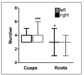
Fig. 3: Distribution of the number of cusps and roots of upper third molars according to side. We determined the number of cusps and roots of right side (n=234) and left side (n = 228) molars. *p < 0.05; ***p < 0.0001.

Fig. 4: Distribution of the measurements of upper third molars according to side. A. Total length. We determined the occlusalapical (OA) length; B. Coronal measurements.We determined the vestibularpalatal (VP), mesiodistal (MD), cervicalocclusalvestibular (COV), and cervicalocclusalpalatal (COP) diameters of right side (n = 234) and left side (n = 228) molars. *p <0.05; **p<0.001.
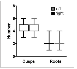
Fig. 5: Distribution of the number of cusps and roots of lower third molars according to side. We determined the number of cusps and roots of right side (n = 304) and left side (n = 195) molars.

Fig. 6: Distribution of the measurements of lower third molars according to side. A. Total length. We determined the occlusalapical (OA) length; B. Coronal measurements. We determined the vestibularlingual (VL), mesiodistal (MD), cervical occlusal vestibular (COV), and cervicalocclusalpalatal (COP)
Upon comparison of the molars from both jaws regardless side, it was observed that the medians for cusps (5) and the COV (7.60 mm), MD (11.26 mm) and OA (17.89 mm) measurements were signifi cantly higher (p< 0.0001) in the lower molars, whereas the number of roots (3) and the COP (7.34 mm) and VP measurements were significantly higher (p < 0.0001) in the upper molars. The dissimilarity and the correlation analysis were performed on arcsinhnormalised dental measure ments abundance levels. Dental measurements could then be clustered according to dental arch and side to determine how closely correlated they were. The dendrogram in Fig. 7 shows the dissimilarity profile of dental measurements grouped to dental arch. The height of each leaf shows dissimilarity of the dental parameter regarding dental arch, i.e., the longer the line of the leaf, the greater the difference. All the resulting clusters were bifolious and each leaf represented the upper and lower arch for each studied dental parameter. There was only one bifolious outlier (leaves 7l and 7u), evidencing the main distinctive feature of this parameter (number of cusps) as regards dental arch. The analysis of the dissimilarity of each clade (dendrogram branch) and its correlation to each dental arch, as merged from the cluster analysis, enabled the assessment of the predictive capability of each dental parameter as regards dental arch. The dental parameters C, MD, OA and COV resulted predictive for lower arch whereas R, VP and COP were predictive for upper arch. The same analysis was performed to assess the predictive capability of the studied parameters for dental side (data not shown). They showed no predictive capability for side in lower molars, although they did in upper molars. In the latter case, C was predictive for left side and R, COP and MD for right side.

Fig. 7: Dendrogram of third molar measurements, roots and cusps.Dissimilarity of the molar morphometric parameters according to dental arch.1 = OA: occlusal apical length; 2 =COV: cervicalocclusalvestibular diameter; 3 = COP: cervicalocclusalpalatal diameter; 4 = MD: mesiodistal diameter; 5 = VP: vestibularpalatal diameter; 6 = R: number of roots;7 = C: number of cusps; u: upper; l: lower.
DISCUSSION
This is the first study updating morphometric data of third molars in recent decades in our country. It is worth noting that the size of the study population of third molars used in this research is the largest ever used. Sample sizes used in previous studies17, 19-21 have always been smaller than one half of the size we used here. In general, most previous studies19-21 have reported morphometric data from molar measurements taken by means of Vernier calipers. In contrast, we report data obtained using digital calipers rather than Verniers, ensuring accuracy and precision of the measurements, as proposed for anthropometric measurements of skeleton remains or cadaveric bones and teeth22. Another interesting feature of our research is the complete descriptive statistical report of the molar measurements, including median, 95% confidence interval and range. In contrast, previous investigations reported only average measurements of some dental para meters of the diameters of right side (n = 304) and left side (n = 195) molars.
molar and no dispersion statistics. However, those averages, although different, are comparable to ours. As a rule, average third molar measurements reported previously17, 19-21 are within the 95% confidence interval of our data reported in this paper. This includes COV, COP, OAdistances, MD and VP, R and C, of both upper and lower third molars. One original contribution of this study is the analysis of the capability of dental morphometric parameters to predict the side or dental arch the molar belongs to. This kind of analysis had not been performed to date. In our research, the study population analyzed is appropriately large to perform such analysis. At present, virtual modeling of bones and teeth is of dental, forensic and anthropological interest7, 16, 18, 23, 24. These specialties require the most accurate and precise data possible. The accuracy and precision of models usually increases when input data are weighted according to their predictive capability18. In this regard, the data reported in this paper will contribute to the development of more accurate mathematical models. However, one limitation of this study to that aim is the number of dental parameters considered. We only studied morphometric parameters, though occlusal topographical parameters such as the perimeter of the molar occlusal view, perimeter of the occlusal surface, number of pits and fissures, cusps height and angles, should also be considered, some of which might reveal higher predictive capability for side or dental arch.
We conclude that despite the morphometric heterogeneity of third molar, there are intrinsic parameters with predictive capability for dental arch and side, but it would be advisable to supplement this study with data from topographic occlusal variables in order to validate the accuracy of their predictive capability.
1. Jokstad A. Methodological challenges in the study of dental occlusion. J Oral Rehabil 2012; 39: 480-488. [ Links ]
2. Thevissen PW, Fieuws S, Willems G. Third molar development: measurements versus scores as age predictor. Arch Oral Biol 2011; 56:1035-1040. [ Links ]
3. Koolstra JH. Dynamics of the human masticatory system. Crit Rev Oral Biol Med 2002; 4:366-376. [ Links ]
4. Sert S, Sahinkesen G, Topcu FT, Eroğlu SE, Oktay EA. Root canal configurations of third molar teeth. A comparison with first and second molars in the Turkish population. Aust Endod J 2011; 37: 109-117. [ Links ]
5. Johan NA, Khamis MF, Abdul Jamal NS, Ahmad B, Mahanani ES. The variability of lower third molar development in Northeast Malaysian population with application to age estimation. J Forensic Odontostomatol 2012; 1: 45-54. [ Links ]
6. Kuzekanani M, Haghani J, Nosrati H. Root and canal morphology of mandibular third molars in an Iranian population. J Dent Res Dent Clin Dent Prospect 2012; 6:85-88. [ Links ]
7. Gaspersic D. Morphometry, scanning electron microscopy and Xray spectral microanalysis of protostylid pits on human lower third molars. Anat Embryol (Berl) 1996; 193: 407-412. [ Links ]
8. Peterkova R, Lesot H, Peterka M. Phylogenetic memory of developing mammalian dentition. J Exp Zool B Mol Dev Evol 2006; 306: 234-250. [ Links ]
9. Esposito M, Coulthard P. Impacted wisdom teeth BMJ Clin Evid. 2008; pii: 1302. [ Links ]
10. Marciani RD. Third molar removal: an overview of indications, imaging, evaluation, and assessment of risk. Oral Maxillofac Surg Clin North Am 2007; 19:113 [ Links ]
11. Choi BJ, Park S, Lee DW, Ohe JY, Kwon YD. Effect of lower third molars on the incidence of mandibular angle and condylar fractures. J Craniofac Surg 2011; 22: 1521-1525. [ Links ]
12. Susarla SM, Dodson TB. Predicting third molar surgery operative time: avalidated model. J Oral Maxillofac Surg 2013; 71:5-13. [ Links ]
13. AlKhatib AR, Rajion ZA, Masudi SM, Hassan R, Townsend GC.Validity and reliability of tooth size and dental arch measurements: a stereo photogrammetric study. Aust Orthod J 2012; 28:22-29. [ Links ]
14. Ganguli A, Bhattacharyya B. Role of morphometry in the identification of teeth: Maxillary permanent molars from an eastern Indian population. Forensic Sci Int 1986; 32: 103-107. [ Links ]
15. Sidow SJ, West LA, Liewehr FR, Loushine RJ.Root canal morphology of human maxillary and mandibular third molars. J Endod 2000; 26:675-678. [ Links ]
16. Klukkert ZS, Dennis JC, M’kirera F, Ungar PS. Dental topographic analysis of the molar teeth of primates. Methods Mol Biol 2012; 915: 145-152.
17. Figun ME, Garino R. Anatomia odontologica funcional y aplicada. Buenos Aires, Argentina. El Ateneo 1978; 244-245, 247-249, 266. [ Links ]
18. Szucs A, Bujtar P, Sandor GK, Barabas J. Finite element analysis of the human mandible to assess the effect of removing an impacted third molar. J Can Dent Assoc 2010; 76:a72. [ Links ]
19. Scott JH, Symons NBB. Introduccion a la anatomia dentaria. Buenos Aires, Argentina. Mundi 1980; Cap.1: 22-28. [ Links ]
20. Riojas Garza, MT. Anatomia dental. Mexico DF, Mexico: El Manual Moderno 2006; 9798, 108-109. [ Links ]
21. Scheid R, Weiss G. Woefel. Anatomia dental. Barcelona, Wolters Kluwer/Lippincott Williams & Wilkins 2012; 153-156. [ Links ]
22. Burns K. Forensic anthropology training manual. New York, NY, USA: Routledge 2012; 153-180. [ Links ]
23. Barone S, Paoli A, Razionale AV. Geometrical modeling of complete dental shapes by using panoramic Xray, digital mouth data and anatomical templates. Comput Med Imaging Graph 2015; 43:112-121. [ Links ]
24. Teshima TL, Patel V, Mainprize JG, Edwards G, Antonyshyn OM. A threedimensional statistical average skull: applica tion of biometric morphing in generating missing anatomy. J Craniofac Surg 2015; 26:1634-1638. [ Links ]

