


Servicios Personalizados
Revista

Articulo
 Inglés (pdf)
 Articulo en XML
 Referencias del artículo
 Enviar articulo por email
Indicadores
-
 Citado por SciELO
Links relacionados
-
 Similares en
SciELO
Similares en
SciELO
Compartir

 Permalink
Acta Odontológica Latinoamericana
versión On-line ISSN 1852-4834
Acta odontol. latinoam. vol.30 no.1 Buenos Aires abr. 2017
ARTÍCULO ORIGINAL
Root surface temperature variation during mechanical removal of root canal filling material. An in vitro study
Variaciones térmicas en la superficie radicular durante la desobturación mecánica del conducto. Estudio in vitro
Martín García-Cuerva1, Lucía Horvath1, Laura Pinasco2, Verónica Ciparelli1, Hernán Tartacovsky1, Ariel Gualtieri3, Ana C Casadoumecq4, Pablo Rodríguez2, Carlos Gonzalez-Zanotto1
1 Universidad de Buenos Aires. Facultad de Odontología. Cátedra de Clínica I de Operatoria
2 Universidad de Buenos Aires. Facultad de Odontología. Cátedra de Endodoncia
3 Universidad de Buenos Aires. Facultad de Odontología, Cátedra de Biofisica
4 Universidad de Buenos Aires. Facultad de Odontología. Cátedra de Fisiología
CORRESPONDENCE
Dr. Martin García Cuerva
Cátedra de Clínica I de Operatoria, Facultad de Odontología, Universidad de Buenos Aires Marcelo T. de Alvear 2142, Piso 9°B,(CP1122AAH) C. A. B. A., Argentina.
E-mail: gc_martin@hotmail.com
ABSTRACT
The aim of this study was to analyze in vitro temperature changes on the outer surface of the dental root during mechanical filling removal procedures.
Thirty recently extracted single-rooted lower premolars were cut transversally at 16 mm from the apex in order to standardize sample length. Endodontic treatment was performed on them. The filling material was subsequently removed using Gates Glidden (G1, G2, G3); Peeso (P1, P2, P3) and PostecPlus FRC (FRC) reamers while temperatures were measured on the outer surface using a digital device with thermocouple at 0, 2, 4, 6, 8, 10 and 15 seconds. Temperatures were compared using repeated measures ANOVA followed by pairwise comparison with Tukey's test.
All reamers caused significant temperature variation between different times (p<0.05). Pairwise comparisons indicated that temperature increased with time for all reamers (p<0.05).
Significant differences in temperature were found between different reamers after 0, 2, 4, 6, 8,10 and 15 seconds (p<0.05). Temperature at the root surface increased considerably. Values higher than 50°C were recorded, the greatest increase from baseline being 16°C. Accordingly, if the procedure were begun at 37°C (physiological temperature), the temperature in the surrounding tissues - cementum, periodontium and bone - would rise to 53°C. An increase in 10°C above body temperature at the root surface may cause lesions in surrounding tissues. While removing filling material, it is essential to cool, control action time and use instruments in perfect condition, all of which may contribute to reducing the heat generated and transmitted to the outer root surface.
Key words: Root canal preparation; Transition temperature; Endodontic.
RESUMEN
El objetivo del presente trabajo fue estudiar los cambios térmicos in vitro en la superficie externa de la raíz del diente, generados durante los procedimientos de desobturación mecánica.
Se utilizaron 30premolares inferiores unirradiculares recientemente extraídos, que fueron seccionados transversalmente a 16 mm del ápice para estandardizar la longitud de las muestras. Se realizó luego su tratamiento endodóntico. Las mediciones de temperatura fueron realizadas mediante un dispositivo digital con termocupla, en la superficie externa a diferentes tiempos (t = 0, 2, 4, 6, 8, 10 y 15 segundos), durante la desobturación con fresas de Gates Glidden (G1, G2, G3); de Peeso (P1, P2, P3) y la correspondiente a PostecPlus FRC (FRC). Los registros de temperatura fueron comparados mediante pruebas de ANOVA de medidas repetidas, seguidas por comparaciones entre pares mediante la prueba de Tukey. Con todas las fresas se encontró una variación significativa de la temperatura entre los diferentes tiempos (p<0.05). Las comparaciones entre pares indicaron que la temperatura se incrementó con el tiempo en todas las fresas (p<0.05). Se detectaron diferencias significativas de temperatura entre diferentes fresas después de 0, 2, 4, 6, 8, 10y 15 segundos (p<0.05).
El aumento de la temperatura de la superficie radicular fue importante ya que los valores registrados superan los 50°C, teniendo en cuenta que el aumento de temperatura mayor fue de 16°C. Si partimos de los 37°C (temperatura fisiológica), la temperatura presente en los tejidos circundantes; cemento, periodonto y hueso; alcanzaría los 53°C. Un aumento de 10°C por encima de la temperatura corporal en la superficie radicular podría causar lesiones en los tejidos circundantes. La utilización de refrigeración, el control del tiempo de acción y el uso de un instrumental en estado óptimo son parámetros ineludibles debido a que los mismos pueden contribuir a disminuir el calor generado y trasmitido hacia la superficie externa de la raíz durante la desobturación.
Palabras clave: Preparación del conducto radicular; Temperatura de transición; Endodoncia.
INTRODUCTION
In teeth with endodontic treatment, crown and root structures are weakened by loss of tissue as a result of previous restorations, caries and preparation for endodontic access. It is therefore important to note that rehabilitating an endodontically treated tooth involves working on a structure that has been diminished both mechanically and biologically. The main reinforcement in an endodontically treated tooth is constituted by its own tissues and anatomical structures, so as a general principle; the selected restoration procedures should preserve as much tissue as possible1. When much of the clinical crown has been lost, the remaining dentin often does not provide sufficient anchorage for a restoration. Such cases call for intraradicular restoration using materials such as posts made from organically based materials and bonded with resin cements to the remaining tooth. Their mechanical behavior is similar to that of dental tissues and thereby improves the distribution of forces 2.
Filling material has to be removed from the canal to provide a smooth bonding surface between wall and anchor, at the same time preserving tooth anatomy. This is done using reamers. It is important to handle and control rotary instrument speed adequately in order to avoid increasing the temperature at the root surface.
Bone tissue is sensitive to temperatures over 47° C (10° higher than body temperature), which may damage microcirculation and connective tissue, cementim, periodontium and alveolar bone as well as causing dentine resorption and chronic immflamation of the periodontim and adjacent bone tissue 3-6. Damage may be reversible if it is limited a and temperature does not exceed 53°C, (alkaline phosphatase denaturing point); however, higher temperatures may cause irreversible bone damage. The mechanisms of such damage are not fully understood. Despite the low thermal conductivity of dentine, it can still transmite heat to the outer surface of the root and tooth-supporting tissues when rotary systems are used during endodontic preparation 7-11 The aim of this study is to analyze temperature changes on the outer surface of the root caused by mechanical procedures for removing filling from a root canal.
MATERIALS AND METHODS
This in vitro study used an experimental design to simulate usual endodontic clinical procedures on 30 single-rooted lower premolars which had been recently extracted by orthodontic indication. Sex, age and reason for extraction were not considered as study variables. Extracted teeth were stored in 0.5% chloramine-T solution at 4 °C.
Inclusion criteria were:
- Straight, single-rooted teeth.
- Conical roots with circumferential diameter 15.5 ± 2.0 mm.
Root length was standardized at 16 mm as measured from apex to crown, at which level it was cut transversally using a diamond disc (KG Sorensen, Brazil) with plentiful cooling. Preoperative periapical radiographs were taken of each tooth for 0.7 second with Skydent Speed E film and New Life Radiology 65KV 8mA Denimed X-ray equiμment by paralleling technique with focus-to-object distance 10 cm, to obtain an image of the longitudinal axis of the tooth. One specialist performed endodontic treatment on all teeth using the ProTaper Universal system (Densply-Maillefer. Ballaigues, Switzerland). Catheterization was performed using a K 10 file, followed by preparation of access using K 10-1520 files (Dentsply Maillefer, Switzerland) and Protaper system S1-S2-Sx files, which were only used on the coronal and middle thirds. Rinses with 10 ml 2.5% sodium hypochlorite were applied between files and canal apical patency was maintained with a No. 10 patency file. Working length was determined by measuring the canal with a K N 15 file. Mechanical preparation was done with ProTaper F1-F2-F3 files for the apical third and ProTaper S1, S2, F1, F2 and F3 files for the middle and coronal thirds. Finally, 17% EDTAC was allowed to act for 5 minutes, simulating the time for which dentin remains in contact with endodontic irrigants, and then rinsed with 10 ml 2.5% sodiumhypochlorite 13,14.
Root canals were dried with standardized absorbent paper points (Dentsply) and filled using hybrid technique with size 30 gutta-percha points (Dentsply-Maillefer), Sealer 26 (Dentsply-Maillefer) and size 15 accessory points, which were thermoplasticized using a size 30 gutta-condensor (Dentsply-Maillefer). Canal openings were sealed with glass ionomer (Vitrebond - 3M. Seefeld, Germany). Samples were stored for 7 days at 37°C and 100% humidity in an oven (Biomerican, model bs615).
Canal preparation and filling removal were performed by one standardized operator in order to reduce bias. Filling material was removed from the canal using Gates Glidden No. 1, 2 and 3 (Dentsply Maillefer, Switzerland), Peeso No. 1, 2 and 3 (Dentsply Maillefer, Switzerland) and Postec Plus FRC 3 system reamers (tapered reamers with 1.3 mm diameter at cervical level and 0.6mm diameter at apical level for size 1, 1.5mm/0.8mm for size 2 and 2mm/1mm for size 3) (Ivoclar Vivadent, Liechtenstein). Each reamer was used to prepare five beds, as intended by the commercial kit. No cooling was used during the procedure. Temperature was measured at baseline (time 0) and at 2, 4, 6, 8, 10 and 15 seconds after the start of filling removal. Temperature variations were measured with a digital device with thermocouple (M890G Temperature Meter, Fig. 1). The sensor was firmly attached to the outer surface of the middle third of the root (Fig. 2).

Fig. 1: Digital device with thermocouple.

Fig. 2: Measuring samples.
Statistical analysis
Results were analyzed statistically. Thirty temperature measurements were recorded for each combination of time and reamer. Mean and standard deviation (SD) were reported for temperatures for each of these groups. Temperature was compared among times for each reamer and among reamers for each time. These comparisons were performed by repeated measures ANOVA, which is appropriate for pairwise data, as in this experimental design15. When ANOVA provided a significant result, pairwise comparisons were performed between groups using Tukey's test. For all tests, results were considered significant when p<0.05. Analyses were performed using the software Infostat version 201316.
RESULTS
Table 1 summarizes the results. Temperature change over time was similar for the different reamers. Repeated measurements ANOVA provided a significant global result (p<0.05). Pairwise comparison with Tukey's test showed that temperature differed significantly among different times, with higher temperatures as time increased. There was only one exception: for Gates 1, pair wise comparisons showed no significant difference between baseline and 2 seconds.
Table 1: Comparison of temperatures among different reamers and times, with repeated measures ANOVA followed by post hoc pairwise comparisons (Tukey's test).
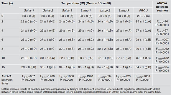
Temperatures were not compared between reamers for baseline (time 0) because all values were equal (23°C). For each subsequent time, significant differences in temperature were found between reamers using repeated measures ANOVA. Specifically, pairwise comparison using Tukey's test showed the following:
- At all times, temperatures with Gates 1 were lower than the rest.
- At two seconds, there was no significant difference among Gates 2, Gates 2, Largo 1, Largo 2 and Largo 3. Temperature with FRC 3 were higher than the rest.
- At four seconds, temperature with Largo 1 was lower than temperature with Gates 2, Gates 3, Largo 2, Largo 3 and FRC 3. Temperature with Largo 3 and FRC 3 was higher than with Gates 2, Gates 3 and Largo 2.
- The results at 6, 8, 10 and 15 seconds were similar. The highest temperature was reached with Largo 3. Temperatures with Gates 3, Largo 2 and FRC 3 were higher than with Gates 2 and Largo 1.
Fig. 3 shows results for baseline and at 8, 10 and 15 seconds.
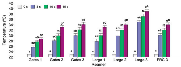
Fig. 3: Temperature reached at different times (0, 8, 10 and 15 seconds) for each reamer (mean + SE). Different lowercase letters indicate significant differences (P<0.05) between times for the same reamer. Different uppercase letters indicate significant differences (P<0.05) between reamers for the same time.
DISCUSSION
The harmful effects of heat on the outer root surface during filling removal procedures are clinically important because an increase greater than 10°C above physiological temperature (37°C) can alter the viability of supporting tissues and cause bone necrosis, cell apoptosis and sometimes, in more severe cases, tooth ankylosis9. Heat generation while removing filling from the canal system can be modified in various ways, including type of instrument used (acuity, sharpness and size), condition of instrument cutting edges, rotation, cutting pressure applied and contact time with tooth structure.
This study found temperature increases of up to approximately 16°C at 15 seconds during canal preparation. Wider reamers were associated to greater heat generation. Manufacturers' protocols for filling removal say that working time should be shorter than 1 minute. Based on our results, we suggest that this time limit should be considerably lower.
Another factor to consider during filling removal is how thick the remaining dentin is in the canal walls. Stripping dentin tissue excessively from the canal walls during preparation with the aim of increasing anchorage and achieving better fit of the element to be bonded is contraindicated. It will not only weaken the walls, but also increase heat transmission outward. Excessive removal of root dentin is known to compromise the root, and preserving root dentin is directly related to root strength 17,18. Thus, knowledge of the internal tooth anatomy contributes to dental practice which is more conservative of tissues, and avoids causing excessive damage to teeth and tooth supporting tissues during preparation. Customizing posts prior to bonding may contribute to reducing the occurrence of irreversible injury. Lubieniecka et al.19 replicated the clinical situation of removing filling from canals during post space preparation and analyzed it with a thermal imaging camera. The effect of cooling was clear in the cervical region of the tooth, where temperature wasvery close to initial temperature reading. The highest temperature on the surface of the root corresponded to the greatest depth that the drill reached. The periapical zone experienced very little or no temperature increase19. Weller et al.20 recorded the highest temperature increase at the most coronal part of the root, which is closely related to larger width of reamers, more gutta-percha in this part of the tooth and thinner dentin walls. Other authors such as Lima Machado and Antoniazzi21 reported higher temperatures for instrumentation of the cervical third than for the middle and apical thirds. Some authors suggest that clinicians should take into account that dentin is considered to be a good thermal insulator, and the thicker it is, the less heat will be transferred to the outer surface of the root22-24. However, depending on their anatomy, not all teeth have a thick layer of dentin, e.g. lower incisors have very thin walls. Care must therefore be exercised to avoid damaging tooth support tissue when remaining dentin is less than 1 mm thick22.
Further studies could consider working length, type and size of reamer used and cooling while preparing root canal. The use of anatomic posts could also be considered, since they are more respectful of tooth anatomy, requiring less thinning of dentin walls and consequently providing treatment that is more conservative of remaining dentin structures.
CONCLUSION
Within the limitations of the present study, the increase in temperature at the root surface during mechanical removal of filling was important, since the values recorded at the outer root surface were high enough to damage tissues surrounding the tooth. Even though temperatures increased to critical values for surrounding tissues, the absolute results of this in vitro study are not directly transferable to real clinical situations, since they would be influenced by periodontal tissues, periodontal blood circulation and the oral environment.
These results suggest the need for further studies to enable current protocols to be adapted.
1. Bertoldi Hepburn. Rehabilitación posendodontica. Capitulo 2, Postura filosófica para la rehabilitation posendodóntica. Buenos Aires, Argentina. Editorial Médica Panamericana S. A. C. F., 2012: 2-19. [ Links ]
2. Menezes MS, Queiroz EC, Campos RE, Martins LRM, et al. Influence of endodontic sealer on fibreglass post bond strength to root dentin. Int Endod J 2008; 41:476-484. [ Links ]
3. Eriksson A, Albrektsson T, Grane B, Mcqueen D. Thermal injury to bone. A vital-microscopic description of heat effects. Int J Oral Surg 1982; 11:115-121. [ Links ]
4. Bhaskar SN, Lilly GE. Intrapulpal temperature during cavity preparation. J Dent Res 1965; 44:644-647. [ Links ]
5. Tjan AH, Abbate MF. Temperature rise at root surface during post -space preparation. J Prosth Dent 1993; 69:41-45. [ Links ]
6. Saunders EM, Saunders WP. The heat generated on the external root surface during post space preparation. Int Endod J 1989; 22:169-173. [ Links ]
7. Eriksson JH, Sundstrom F. Temperature rise at root surface during root canal preparation a possible cause of damage to tooth and periodontal tissue. Swed Dent J 1984; 8:217-223. [ Links ]
8. Cohen BI, Deutsch AS, Musikant BL. Effect of power settings on temperature change at the root surface when using a Holmium YAG laser in enlarging the root canal. J Endod 1996; 22:596-599. [ Links ]
9. Eriksson AR, Albrektsson T. Temperature threshold levels for heat-induced bone tissue injury: A vital-microscopic study in the rabbit. J Prosthet Dent 1983; 50:101-107. [ Links ]
10. Kilic K, Er O, Kilinc HI, Aslan T, et al. Infrared thermographic comparison of temperature increases on the root surface during dowel space preparations using circular versus oval fiber dowel systems. J Prosthodont 2013; 22:203-207. [ Links ]
11. Ratih DN, Palamara JEA, Messer HH. Temperature change, dentinal fluid flow and cuspal displacement during resin composite restoration. J Oral Rehabil 2007; 34:693-701. [ Links ]
12. Lipski M, Mrozek J, Drozdzik A. Influence of water cooling on root surface temperature generated during post space preparation. J Endod 2010; 36:713-716. [ Links ]
13. Cunha RS, De Martin AS, Barros PP, da Silva FM, et al. In vitro evaluation of the cleansing working time and analysis of the amount of gutta-percha or Resilon remnants in the root canal walls after instrumentation for endodontic retreatment. J Endod 2007; 33:1426-1428. [ Links ]
14. Galafassi D, Colucci V, Cecchin D, Scatena C, et al. Effect of Endodontic Irrigants on Microtensile Bond Strength to Dentin After Thermocycling and Long-Term Water Storage. J Dent (Tehran) 2013; 10:426-435. [ Links ]
15. Davis C. Normal-Theory Methods: Repeated Measures ANOVA. En: Statistical methods for the analysis of repeated measurements, 1ra ed. New York: Springer-Verlag, 2002: 103-116. [ Links ]
16. Di Rienzo JA, Casanoves F, Balzarini MG, Gonzalez L, et al. InfoStat versión 2013p, Grupo InfoStat, FCA, Universidad Nacional de Córdoba, Argentina. 2013. [ Links ]
17. Lertchirakarn V, Timyam A, Messer HH. Effects of root canal sealers on vertical root fracture resistance of endodon-tically treated teeth. J Endod 2002; 28:217-219. [ Links ]
18. Fukui Y, Komada W, Yoshida K, Otake S, et al. Effect of reinforcement with resin composite on fracture strength of structurally compromised roots. Dent Mater J 2009; 28: 602-609. [ Links ]
19. Lubieniecka J, Lukasiewicks J, Bozyk J, Kleinrok J. The evaluation of increase and distribution of temperature during the dental drilling using a thermal imaging camera. FLIR technical series. Application note for research and science. 2011: 1-7. [ Links ]
20. Weller RN, Kimbrough WF, Anderson RW. Root surface temperatures produced during post space preparation. J Endod 1996,22: 304-307. [ Links ]
21. Lima Machado ML, Antoniazzi JH. In Vitro evaluation of the temperature achieved by Gates-Glidden. Largo and modified largo drills during chemo-surgical preparation of root canals. Ecler endond. Vol 1 n3 Sao Paulo, 1999. [ Links ]
22. Kwon SJ, Park YJ, Jun SH, Ahn JS, et al. Thermal irritation of teeth during dental treatment procedures. Restor Dent Endod 2013; 38: 105-112. [ Links ]
23. Ottl P, Lauer HC. Temperature response in the pulpal chamber during ultrahigh-speed tooth preparation with diamond burs of different grit. J Prosthet Dent 1998; 80:12-19. [ Links ]
24. Dominici JT, Clark S, Scheetz J, Eleazer PD. Analysis of heat generation using ultrasonic vibration post removal. J Endod 2005; 31:301-303. [ Links ]

